Alternative Interventional/Surgical Methods for the in-vivo - PowerPoint PPT Presentation
Alternative Interventional/Surgical Methods for the in-vivo Thrombogenicity Test Kent Grove MS, HT (763)951-8097 kgrove@apsemail.com Traditional Method Canines are the preferred animal species (n=2). The animals are un-heparinized.
Alternative Interventional/Surgical Methods for the in-vivo Thrombogenicity Test Kent Grove MS, HT (763)951-8097 kgrove@apsemail.com
Traditional Method • Canines are the preferred animal species (n=2). • The animals are un-heparinized. • A percutaneous stick or surgical cut down is performed to gain access to the treatment site. • Equal lengths of both test and control devices (~10 cm) are inserted into alternating Right and Left Jugular Veins. • The devices secured to the surrounding tissue to prevent movement. • The devices are implanted for a total of 4-hours. • Just prior to euthanasia, a bolus injection of heparin is administered to help prevent post mortem clotting. • Devices are explanted and evaluated for the presence of thrombus in comparison to the control device.
Traditional Method (Key Concept) • Reduction in blood flow = Increased probability of thrombus formation. • Variables that can affect blood flow • Treatment site access and securing the device to prevent movement during the 4-hour period. • Animal positioning. • Location of valves within the treatment sites. • Vessel Size and Device Placement.
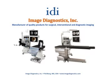
Lets Take a Closer Look • The use of fluoroscopic imaging can help to reduce a number of variables that the current model can overlook.
Animal Positioning • The positioning of the animal throughout the study can have a dramatic impact on the blood flow around the device.
Blood Valves • Variability in the locations of blood valves can vary from one animal to the next. • A difference can be seen when comparing right and left jugular veins in the same animal. • Placement of a device through a blood valve(s) can reduce the blood flow in that particular treatment site. • This can cause variability amongst the treatment sites.
Vessel Size and Device Placement • Size of the vessel in relation to the device can have a impact on thrombus formation. • The size and shape of the device should be considered prior to deployment. • Patency and proper deployment of the device must be evaluated immediately post implant with the use of fluoroscopy. • If the Device is in contact with the vessel wall during the 4-hour indwelling period, there is a higher probability of thrombus formation.
Consider using both the Jugular and Iliac veins • The Iliac veins provide another treatment site for both test and control device. • The Iliac veins have adequate blood flow while the animal is on the table. • This design allows for an evaluation of one additional test and control device in each animal on study. • This helps to reduce the potential of receiving conflicting results.
Jugular and Iliac Method Considerations • There are many procedural related variables that can affect blood flow. • The use of fluoroscopic imaging can help to reduce these variables. • Vessel size should be evaluated prior to implantation. * As a general rule, any device larger than 10 Fr should be evaluated in a sheep. • Proper evaluation of the device deployment and patency should be evaluated with the use of angiography. • This method should considered for the evaluation of devices such as dilators, sheaths, guidewires and standard catheters.
Swine Atrial Model (surgical approach) • Two swine are subjected to bilateral atrial implants. • A sternotomy is performed, a sheath is placed, and the test and control devices are deployed into the right and left atrium. • The use of contrast and fluoroscopic imaging must be used to ensure the proper deployment of the devices prior to removing the sheaths. • This design can be used without Heparin.
Considerations for the Swine Atrial Model • This devices can be implanted without the use of Heparin. • Device Recommendation: Balloon Catheters, Mapping Catheters and Larger devices.
Interventional Thrombogenicity Test • Canine, Sheep or Swine can be used. • Clinical relevance (intended implant location, venous verses arterial blood flow), vessel size, blood flow, size and shape of the device should be considered with the design. • A surgical cut down or percutaneous stick may be used to gain access to the vascular system (example: femoral vein access) • Heparin is administered to maintain an Activated Clotting Time (ACT) level > 250 seconds during the device placement procedure.
Interventional Thrombogenicity Test • Devices are deployed under fluoroscopic guidance. • Fluoroscopic images are obtained with the use of contrast to ensure proper placement and blood flow around the device. • ACT’s are taken until the animal approaches baseline and then the four hour indwelling period begins. • I will give a few examples of recommended deployment sites, but this design is very robust, minimally invasive, and the model is completely customizable to the device.
Interventional Thrombogenicity Test • By far the best design. • Minimally invasive. • Devices are deployed under fluoroscopic guidance. • Fluoroscopic images are obtained with the use of contrast to ensure proper placement and blood flow around the device. • The design is customized to the device.
Thrombus Evaluation • Take Pictures!!!
Thrombus Evaluation Scores
Interpretation of the Results
Thank You!!!
Recommend
More recommend
Explore More Topics
Stay informed with curated content and fresh updates.

