
- einstein. 2016;14(3):439-42
MEDICAL DEVELOPMENTS
This content is licensed under a Creative Commons Attribution 4.0 International License.
A pictorial presentation of 3.0 Chicago Classification for esophageal motility disorders
Apresentação em imagens da Classificação de Chicago versão 3.0 das doenças da motilidade esofagiana
Fernando Augusto Herbella1, Priscila Rodrigues Armijo1, Marco Giuseppe Patti2
ABSTRACT
High resolution manometry changed several esophageal motility
- paradigms. The 3.0 Chicago Classification defined manometric criteria
for named esophageal motility disorders. We present a pictorial atlas of motility disorders. Achalasia types, esophagogastric junction obstruction, absent contractility, distal esophageal spasm, hypercontractile esophagus (jackhammer), ineffective esophageal motility, and fragmented peristalsis are depicted with high-resolution manometry plots. Keywords: Manometry/methods; Esophageal motility disorders; Esophageal achalasia/classification
RESUMO
A manometria de alta resolução mudou vários paradigmas da motilidade
- digestiva. A Classificação de Chicago, na versão 3.0, definiu critérios
manométricos para as doenças da motilidade esofagiana. O presente artigo é um atlas das dismotilidades descritas. Tipos de acalásia,
- bstrução ao nível da junção esofagogástrica, contrações ausentes,
espasmo esofagiano distal, esôfago hipercontrátil, motilidade esofagiana ineficaz e peristalse fragmentada são mostradas em traçados de manometria de alta resolução. Descritores: Manometria/métodos; Transtornos da motilidade esofágica; Acalásia esofágica/classificação
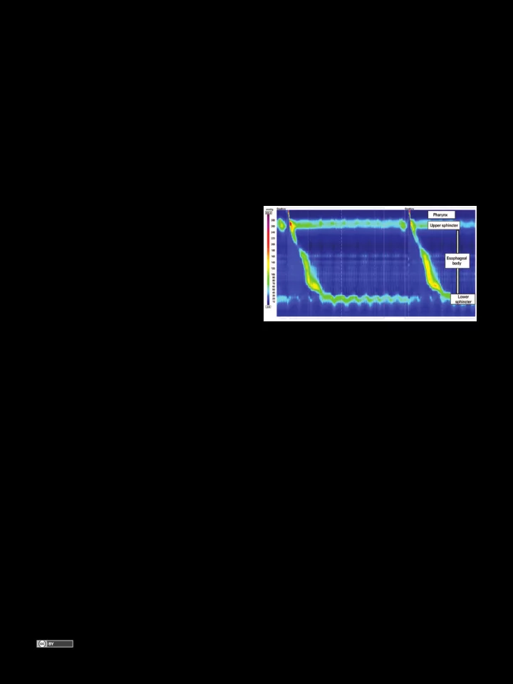
INTRODUCTION High resolution manometry (Figure 1) has clear and inherent advantages over conventional manometry, despite its higher cost.(1) High resolution manometry detailed analysis of esophageal peristalsis changed several esophageal motility paradigms, including new manometric
parameters and different classification for named “motility disorders based on pressure topography”, the Chicago classification,(2) which was recently revised.(3) We present a pictorial atlas of the motility disorders according to the 3.0 Chicago Classification with high- resolution plots.
Achalasia
Chicago Classification divided achalasia into three subtypes according to esophageal pressurization(4) (Figure 2). Type I is characterized by 100% failed contractions and no esophageal pressurization; type II has panesophageal pressurization in at least 20%
- f swallows; and type III is defined by the presence of
preserved fragments of distal peristalsis or premature contractions for at least 20% of the swallows.(3) This classification may be applied to Chagas’ disease esophagopathy as well, although type III is rarely, if ever, seem.(5)
Figure 1. Normal high resolution manometry
1 Escola Paulista de Medicina, Universidade Federal de São Paulo, São Paulo, SP
, Brazil.
2 University of Chicago, Chicago, Illinois, Estados Unidos.
Corresponding author: Fernando Augusto Herbella – Rua Diogo de Faria, 1.087, room 301 – Vila Clementino – Zip code: 04037-003 – São Paulo, SP , Brazil – Phone: (55 11) 3926-7610 E-mail: herbella.dcir@epm.br Received on: July 24, 2015 – Accepted on: Nov 4, 2015 DOI: 10.1590/S1679-45082016MD3444