SLIDE 1
THE UNIVERSITY OF VIRGINIA DEPARTMENT OF OTOLARYNGOLOGY – HEAD & NECK SURGERY PRESENTS
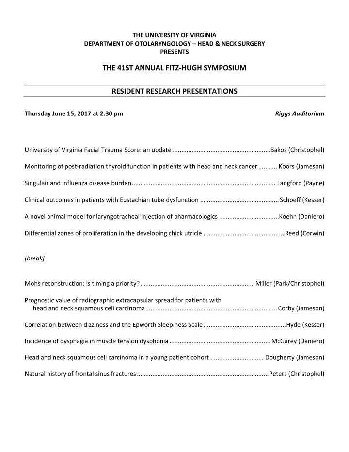
THE 41ST ANNUAL FITZ-HUGH SYMPOSIUM RESIDENT RESEARCH PRESENTATIONS
Thursday June 15, 2017 at 2:30 pm Riggs Auditorium University of Virginia Facial Trauma Score: an update .........................................................Bakos (Christophel) Monitoring of post-radiation thyroid function in patients with head and neck cancer ........... Koors (Jameson) Singulair and influenza disease burden .................................................................................... Langford (Payne) Clinical outcomes in patients with Eustachian tube dysfunction .............................................. Schoeff (Kesser) A novel animal model for laryngotracheal injection of pharmacologics ...................................Koehn (Daniero) Differential zones of proliferation in the developing chick utricle ............................................... Reed (Corwin) [break] Mohs reconstruction: is timing a priority? ................................................................... Miller (Park/Christophel) Prognostic value of radiographic extracapsular spread for patients with head and neck squamous cell carcinoma ............................................................................. Corby (Jameson) Correlation between dizziness and the Epworth Sleepiness Scale ................................................ Hyde (Kesser) Incidence of dysphagia in muscle tension dysphonia ........................................................... McGarey (Daniero) Head and neck squamous cell carcinoma in a young patient cohort ............................... Dougherty (Jameson) Natural history of frontal sinus fractures ............................................................................. Peters (Christophel)
SLIDE 2 2
RESIDENT RESEARCH ABSTRACTS
University of Virginia Facial Trauma Score: an update
Stephen Bakos MD PhD; Garrett Casale MD; Brian Fishero MD; Jared Christophel MD MPH Objective: To correlate applied kinetic energy to facial trauma severity using the University of Virginia facial trauma score (UVAFTS). Design: Creation of facial bony trauma using 8 cadaver heads in a reproducible manner with recording of kinetic energy applied, distance of facial crush, and dentition. Heads then underwent fine cut CT scan. Facial bony trauma graded using the UVAFTS by two raters. Severity of the facial bony trauma, based on the UVAFTS, was compared to kinetic energy applied to the heads. Setting: Center for applied biomechanics and cadaveric dissection laboratory Participants: Otolaryngology residents, otolaryngology attending physician, and staff from the center for applied biomechanics. Results: The UVAFTS correlated with kinetic energy applied to the cadaver heads. Dentition appeared to have a protective role in facial trauma. Inter-rater reliability of the UVAFTS was similar between the two raters. Conclusions: The applied kinetic energy on cadaveric heads correlated to severity of bony facial trauma based on the
- UVAFTS. This novel trauma classification may become an easy and valuable tool to rate severity of facial bony
- trauma. Additional studies are needed to further evaluate the relationship between kinetic energy applied and
severity of facial bony trauma using the UVAFTS.
Monitoring of post-radiation thyroid function in patients with head and neck cancer
Paul Koors MD; Abel David MS4; Mark Jameson MD PhD Objective: To review the head and neck cancer patients treated at UVA in 2014 and determine whether or not patients underwent recommended thyroid hormone screening in 2015 and 2016. A secondary aim was to determine the incidence of post-radiation hypothyroidism and its association with laterality of neck radiation and institution performing the treatment. Design: Retrospective case review. Setting: Tertiary care center. Patients: All head and neck cancer patients treated with radiation to cervical nodes in 2014 were included in the study. Main Outcome Measures: Presence or absence of TSH and FT4 studies and results when performed. Ipsilateral vs bilateral neck radiation was noted as was the center that delivered the radiation. Results: 74 patients underwent radiation therapy to cervical nodes in 2014 and did not meet exclusion criteria. 54%
- f these patients (40/74) underwent regular TSH monitoring. For patients treated at UVA, regular monitoring
- ccurred in 63.5% (33/52), while monitoring was documented in only 31.8% of patients treated elsewhere (7/22).
50% of monitored patients became hypothyroid, 45.5% of patients treated at UVA vs. 57.1% of patients treated elsewhere (p=0.5738). 26.7% and 60% of patients treated with unilateral and bilateral radiation became hypothyroid, respectively (p=0.0410). Conclusion: 45.9% of patients who were seen at UVA for head and neck cancer in 2014 and underwent cervical RT did not receive regular TSH monitoring. In order to improve survivorship management, measures are needed to ensure compliance with this national guideline.
SLIDE 3 3
Singulair and influenza disease burden
Brian Langford MD; Abel David MS4; Thomas Braciale MD PhD; Spencer Payne MD Objective: To investigate whether Montelukast therapy is associated with decreased influenza infection severity. Design: Retrospective cohort study. Setting: Tertiary care academic medical center. Participants: All patients in the Clinical Data Repository (CDR) database 2013-2016. Main Outcome Measures: Influenza infection, influenza pneumonia, hospital admission, ICU admission, and need for an emergent airway. Results: 6004 patients in the CDR had diagnoses of influenza infection. 2387 patients were on Montelukast, 28 of whom were diagnosed with influenza infection (1.2%). Odds ratios were calculated. Admission was more likely for flu (164), flu pneumonia (31.2), asthma flu (47.8), and asthma flu pneumonia (2.25) in patients on Montelukast. ICU admission was more likely for flu (8.1), flu pneumonia (5.7), and asthma flu (5.3), but less likely for asthma flu pneumonia (0.33) in patients on Montelukast. An emergent airway was not required for any of the 28 patients on Montelukast with flu. Conclusions: There is no conclusive evidence that Montelukast usage is associated with decreased disease severity in patients with influenza infection.
Clinical outcomes in patients with Eustachian tube dysfunction
Stephen Schoeff MD; Candice Kremer BS; Brian Hernandez MD; Bradley Kesser MD Objectives: 1) Describe the clinical course of Eustachian tube dysfunction. 2) Identify trends associated with disease progression Study Design: Retrospective cohort study of patients seen at the neurotology division of a tertiary care center’s
- tolaryngology department with a diagnosis of Eustachian tube dysfunction over a 7.5 year period starting in January
2009. Outcome Measures: The primary outcome measure was surgery free survival. Additional outcome measures included audiologic outcomes, use of hearing aids, and number of surgeries. Results: 255 patients were seen in 1361 encounters for an average duration of 43 months. A total of 325 lifetime surgeries were performed on 152 patients. The majority (82%) had bilateral disease. Surgery free survival did not differ significantly when comparing use of intranasal steroids (p=0.058), CPAP use (p=0.19), smoking (p=0.482), and allergic rhinitis (p=0.587). Speech discrimination score significantly worsened in both ears over the course of follow
- up. Laterality and gender were not predictive of cholesteatoma development, need for surgery, and need for hearing
- aids. Age at first surgery was correlated with age at hearing aid acquisition (0.948, p=0.000) and inversely correlated
with total number of surgeries (-0.262, p=0.002). Number of surgeries and number of visits over the study period were positively correlated (0.181, p=0.004). Age of presentation was significantly lower among patients requiring surgery than those not requiring surgery (p=0.001). Conclusion: Eustachian tube dysfunction remains a challenging disease to quantify with a substantial burden on
- adults. Our data add to the existing data in further clarifying the course of the disease and its significant outcomes.
SLIDE 4 4
A novel animal model for laryngotracheal injection of pharmacologics
Heather Koehn MD; Kevin Carlson MS4; Patrick Cottler PhD; James Daniero MD MS Objective: Hillel et al. (2014) have developed a murine model for laryngotracheal stenosis. However, there has not been a corresponding therapeutic model for intralesional injection of laryngotracheal stenosis. Our objective is to design a novel small-animal model for therapeutic injection of pharmacologic agents into the subglottic/tracheal mucosa. Design: IACUC approval was obtained for the use of a murine model. After sedation induced, microlaryngoscopy was attempted, followed by a tracheal cut down exposing the thyroid cartilage to the 3rd tracheal ring. Using a 30-gauge needle a solution of methylene blue and Triamcinolone were injected in the tracheal mucosa inferior to the cricoid
- cartilage. After sacrifice of the animal, tracheal section was harvested for histologic assessment.
Subjects: Two C57BL/6 mice. Intervention: Triamcinolone was selected as the pharmacologic agent for injection due to its wide-spread use in the treatment of laryngotracheal stenosis as well as its molecular properties of fat solubility for histologic evaluation. Results: Adequate endoscopic view was unable to be obtained. The animals experienced respiratory distress as a result of manipulation of the airways using microlaryngoscopy and associated positioning. Without a flexible endoscope, appropriate for the murine airway, the use of the cut down procedure is necessary to visualize the cricoid and perform injection of steroid into tracheal mucosa. Conclusions: The use of the cut down procedure demonstrated access to cricoid for infiltration of triamcinolone in the tracheal mucosa. An endoscope of narrow caliber would be necessary for attaining adequate endoscopic visualization for injection.
Differential zones of proliferation in the developing chick utricle
Robert Reed MD; Mark Rudolf MSE; Jeffrey Corwin PhD Objective: Characterize patterns of embryologic growth in the developing chick utricle. Design: Biological study. Subjects: White Leghorn Chick eggs age E7 to E14. Intervention: 50μg of EDU was injected into E7 eggs via a direct egg injection technique vs. intravenously in E14 eggs. Utricles were subsequently harvested at later time points ranging from 2 hours to 5 days. The utricles were prepared with the click-it reaction and stained with Hoescht (DNA stain) and Myo7A (hair cell marker) via
- immunohistochemistry. The subjects were then analyzed under high power light microscopy.
Results: The utricle was divided into 3 separate zones based on identifiable landmarks and EDU expression: a peristriolar area, a central area, and a medial area. A statistically significant higher level of EDU expression along the medial area compared to striolar area in E9 chicks (p = 0.006) and a high level of EDU expression in the medial area compared to the central region in E14 chicks (p = 0.013) was found. Conclusions: EDU injections in chicks, via direct injections in eggs <E12 or IV injections in eggs >E12 is a feasible way to track cell cycle progression and proliferation. EDU tracing reveals substantial interstitial and appositional growth patterns are present in the chick utricle with preliminary data suggesting the presence of medial appositional growth zone overlying the innervating nerve bundle.
SLIDE 5 5
Mohs reconstruction: is timing a priority?
Matthew Miller MD; Abel David MS4; James McLean MS4; Stephen Park MD; Jared Christophel MD MPH Objective: The timing of Mohs reconstructive surgery is influenced by different factors including surgeon and patient schedules, complexity of defect, and operating room availability. Recent work found that delaying reconstruction by more than two days increases postoperative complication rate. We performed this study to review the outcomes of Mohs Micrographic Surgery (MMS) reconstruction with respect to patient- and surgery-specific variables, especially timing of repair. Design and Participants: This was a retrospective, single-institution cohort study from January 2012 to March 2017. All patients reviewed in the series underwent Mohs reconstructive surgery by one of the two senior authors. No patients had to be excluded for inadequate follow up or incomplete medical records. Primary outcome was post-
- perative complications including hematoma, infection, dehiscence, and partial or full graft or flap loss.
Results: A total of 633 defects in 591 patients were identified over the 5-year period. Reconstructions occurred from <24 hours to 32 days after MMS, with 36.2% delayed greater than 48 hours. Patient-specific variables reviewed included comorbidities, age, smoking status, and use of anticoagulant or antiplatelet medications. Surgery-specific variables analyzed included location and size of defect, time interval between MMS and reconstruction, and reconstructive modalities. Single variable analysis was performed and this was used to form a multivariable binary logistic regression model demonstrating that smoking status, defect size, full-thickness defects, interpolated flaps with cartilage grafting, and composite grafts were associated with an increased risk of postoperative complications (OR 2.50, P = 0.01 [95% CI 1.31-4.78]; Exp(B) 1.04, P = 0.01 [95% CI 1.01-1.06]; OR 1.56, P = 0.02 [95% CI 1.08-2.25]; OR = 8.05, P < 0.001 [95% CI 2.63-24.60]; OR = 6.06, P = 0.001 [95% CI 2.18-16.84], respectively). Conclusions and Relevance: This review demonstrates no association between timing of Mohs reconstructive surgery and complications. Variables associated with an increased risk of postoperative complications include smoking status, size of the defect, full-thickness defects, interpolated flaps with cartilage grafting, and the use of composite grafts.
Prognostic value of radiographic extracapsular spread for patients with head and neck squamous cell carcinoma
Margeaux Corby MD; Brian Hughley MD; Prashant Raghavan MD; Sugoto Mukherjee MD; Henry Frierson MD; Mark Jameson MD PhD Objectives: To evaluate the association of radiographic extracapsular spread with recurrence-free survival. Study Design: Retrospective cohort study Methods: A chart review was performed of patients who underwent neck dissection for HNSCC over a 5-year period at a single institution. Included patients had a preoperative contrasted neck CT scan that had been reviewed independently by 2 neuroradiologists and histological sectioning of all positive nodes reviewed by a surgical pathologist blinded to radiologist results. The presence of radiologic ECS was rated as 0 (absent), 1 (possible), 2 (probable), or 3 (present) in each neck. The extent of histologic ECS was scored as 0-4 using a standard scale ranging from no capsular invasion through nodal obliteration. Kaplan-Meier survival analysis was used to compare radiographic and pathologic groups and recurrence disease-free survival (RFS). Results: In patients with pathologic nodal disease, there was significant difference in RFS between those with nodal disease limited to the nodal capsule and those any extranodal spread (p=.002). There were no significant differences in RFS between groups based on radiographic ECS. Conclusions: In our cohort of patients who underwent primary surgery, any pathologic ECS was associated with poorer RFS. However, there was no correlation between radiographic ECS and RFS. Evidence of extracapsular spread
- n CT does not appear to be a reliable prognostic indicator of RFS in our population.
SLIDE 6
6
Correlation between dizziness and the Epworth Sleepiness Scale
Justin Hyde MD; Garrett Casale MD; Bradley Kesser MD Objective: To correlate daytime sleepiness symptoms via the Epworth Sleepiness Scale (ESS) with complaints of dizziness in patients presenting to otology clinic. Design: Survey with statistical analysis Methods: The ESS was administered to patients presenting to UVA otology clinic over a period of 6 weeks. We then compared their ESS scores with various parameters and demographics including: complaint of dizziness that day in clinic, absolute history of dizziness, men versus women, and adults versus children. Finally we ran 2-tailed T-tests to evaluate for statistical significance. Results: The study included a total of 109 patients who completed the survey. None of the comparisons of interest resulted in statistical significance when compared to their ESS scores: patients with same-day dizziness vs those without (p-value 0.14) and those with a history of dizziness compared to those without (p-value 0.58). There was no demographic difference as men were equivalent to women (p-value 0.15), and adults were equivalent to children (p- value 0.46). Conclusions: Among respondents to the ESS survey, there were no statistically significance differences between patients presenting with dizziness and their sleepiness symptoms within the parameters in question. A larger sample size may be needed for additional analysis. Future directions could include evaluating dizzy symptoms in those with Obstructive Sleep Apnea (OSA) who are treated with CPAP vs those with OSA who are untreated.
Incidence of dysphagia in muscle tension dysphonia
Patrick McGarey MD; Michael Freeman, MD; James Daniero MD MS Objective: To characterize the incidence and nature of reported dysphagia among patients presenting with muscle tension dysphonia. Study Design: Retrospective chart review from October 2014 to December 2015. Setting: Voice and Swallowing Clinic of a tertiary academic medical center. Subjects and Methods: Patients presenting for dysphonia at the University of Virginia Voice and Swallowing Clinic were evaluated by a single fellowship-trained laryngologist. After including Patients who were given a diagnosis of muscle tension dysphonia were included. After excluding patients with pre-existing anatomic causes of dysphagia or prior head and neck cancer, 38 patients were suitable for analysis. Clinical data and patient reported outcomes measures (Voice Handicap Index-10, Eating Assessment Tool-10, Clinical COPD Questionnaire) were reviewed. Results: The co-incidence of patient reported dysphagia among patients with muscle tension dysphonia was 17/38 (44.7%). Among the entire cohort, 25/38 (65.8%) patients had EAT- dysphagia or had abnormal EAT- dysphagia (17.1 vs 10.5 with only dysphonia, p = 0.03). Patients who reported dysphagia also had significantly higher Clinical COPD Questionnaire scores (2.2 vs 0.8 for dysphonia only cohort, p=0.002). Conclusions: The incidence of reported dysphagia among patients with MTD is roughly 50%. Patients who reported dysphagia had significantly higher voice impairment and higher more often reported dyspnea. Further prospective studies are needed to clarify the underlying pathophysiology of dysphagia related to muscle tension, and to investigate diagnostic and therapeutic paradigms.
SLIDE 7
7
Head and neck squamous cell carcinoma in a young patient cohort
William Dougherty MD; Michael Dougherty MS2; Mark Jameson MD PhD Objective: Head and neck squamous cell carcinoma (HNSCC) is rare in patients under 40 years old. However, it is thought to be a more aggressive disease in this age group, and perhaps increasing in incidence. We sought to compare young patients treated at UVA for HNSCC to our older population. Design & Patients: A chart review was performed on all patients under 40 treated at UVA for p16- HNSCC (n=37). A randomized stage and subsite matched cohort aged 55-65 was analyzed for comparison (n=74). SEER9 data were evaluated to note changes in incidence of HNSCC in young patients from 1973-2014. Results: 55% of young patients were never smokers (vs. 12% of older patients, p<0.001) and 41% were female (vs. 22% of older patients, p=0.045). 73% of the young patients had oral tongue cancers. The young patients had better 3- year overall survival than the older group (94% vs. 80%, respectively) but worse 3-year similar disease-free survival (63% vx. 77%, respectively). 56% of young patients had tumors with adverse histologic features (vs. 39% of older patients, p=0.10). A review of the SEER database suggests an increase in tongue cancer amongst young patients, but this trend may be confounded by inclusion of HPV-related tongue base cancers. Conclusions: Despite its limitations, this study supports the idea that young patients who develop HNSCC tend to have more aggressive disease, which may recur earlier but in patients who better tolerate repeated treatment, presumably due to better overall health and nutritional status.
Natural history of frontal sinus fractures
Daniel Peters MD; Jared Christophel MD MPH Objective: To characterize the natural healing process of displaced anterior and/or posterior table frontal sinus fractures. Design: Case series Setting: Ambulatory care at a tertiary care center Patients: A search was performed for patients diagnosed with a frontal sinus fracture in the outpatient setting by our Facial Plastic and Reconstructive Surgery division between 2013 and 2017. Patients were included in the series if they were found to have a displaced anterior and/or posterior table fracture, were managed with observation, and had radiographic follow-up at least three months after the initial diagnosis. Twenty-three patients were identified as having frontal sinus fractures. Seven patients met inclusion criteria. One patient was excluded on the basis of inadequate duration of radiographic follow-up. Six patients were excluded based on lack of radiographic follow-up. Nine patients were excluded based on undergoing operative intervention. Three patients were excluded because the fractures were non-displaced. Main Outcome Measure: There were no outcome measures as the objective of the study was to characterize a population. Results: Seven patients met inclusion criteria. All patients achieved aeration of the frontal sinuses. All patients demonstrated regrowth of bone. All patients demonstrated improvement in bony contour. No patients developed a cerebrospinal fluid leak. No patients developed a mucocele. Conclusion: Observation is a safe management choice for patients with a displaced fracture of the anterior and/or posterior table of the frontal sinus.