
SLIDE 1
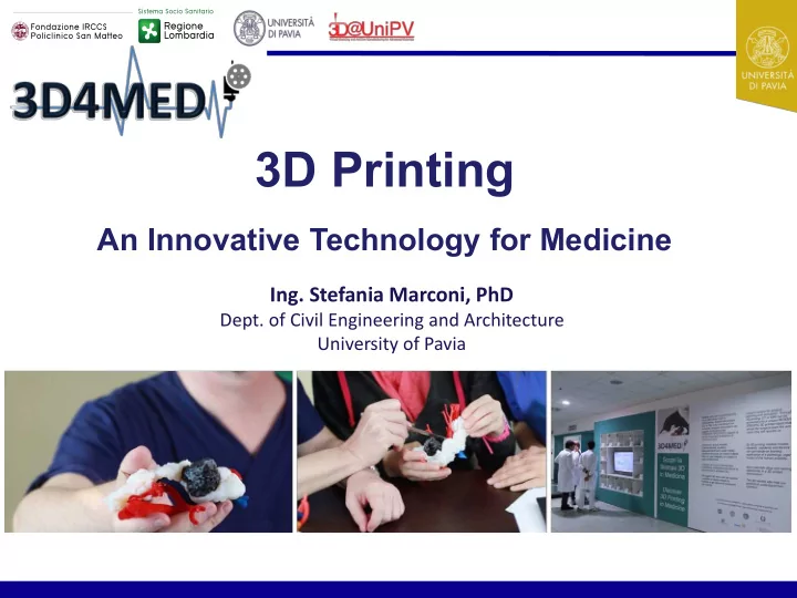
3D Printing
An Innovative Technology for Medicine
- Ing. Stefania Marconi, PhD
- Dept. of Civil Engineering and Architecture
University of Pavia
3D Printing An Innovative Technology for Medicine Ing. Stefania - - PowerPoint PPT Presentation
3D Printing An Innovative Technology for Medicine Ing. Stefania Marconi, PhD Dept. of Civil Engineering and Architecture University of Pavia 3D4MED: the Lab 3D4MED is the first Clinical 3D Printing Lab in Italy and one of the first worldwide.
University of Pavia
It is located at the DEA building of IRCCS Policlinico San Matteo of Pavia and it has a strategic position to improve its visibility (to disseminate the new proposed service) and centrality (to facilitate the collaboration between surgeons and engineers). DEA Building 0 floor – tower B 3D4MED Lab
3D4MED is equipped with all the hardware and software necessary to perform the entire process, from medical images elaboration to the production of the 3D printed model. We are equipped with: ➢ Image elaboration and segmentation software; ➢ Software for virtual models’ manipulation; ➢ 3D printers management software; ➢ Professional 3D printers; ➢ Post-processing instrumentation.
We transform medical images (CT & MRI) into a 3D printed object to hold in your hands!
Virtual Model Slicing 3D Printed Model Thanks to innovative 3D printing technologies, we produce three-dimentional replicas of each patient’s unique anatomy, showing all the surgical relevant structures accurately.
The Lab is equipped with different 3D printing technologies:
3DSystems ProJet 460 Plus
➢ Binder jetting technology; ➢ High precision (100 μm) and low production times and costs; ➢ 2.8 milion colors; ➢ Big/small models with fine details, especially bone structures;
ObJet 260 Connex 3
➢ PolyJet printer with photopolymer resins; ➢ Different colors & materials (deformable and transparent); ➢ Big/small models with fine details (20 μm); ➢ 3D printed models of the abdominal cavity;
FORM 2 Desktop SLA
➢ Printer with photopolymer resins; ➢ High precision (100/50/25 μm); ➢ 4 different transparent materials (1 deformable); ➢ Medium/small (vascular) models with fine details;
3NTR A4v2
➢ Professional FDM printer; ➢ Dual bowden extruder (water cooled extruders up to 410°C);
LeapFrog Creatr HS
➢ FDM printer; ➢ Dual bowden extruder; ➢ Suitable for relatively high speeds printing of large objects;
LeapFrog Creatr Dual Extruder
➢ FDM printer; ➢ Dual direct extruder; ➢ Suitable for low modulus filaments printing;
3NTR A4v3
➢ Professional FDM printer; ➢ Triple bowden extruder (water cooled extruders up to 410°C); ➢ Hot chamber (up to 70°C);
Renishaw AM 400
➢ SLM metal 3D printer; ➢ Flexible AM for professional use in a wide range of metals; ➢ 400W optical system with a reduced beam diameter of 70 μm; ➢ Available metal powders: Titanium, Aluminium, Cobalt chromium, Stainless steel, Nickel alloys.
Instrumentation for post-processing treatments:
➢ Heat treatments oven; ➢ Sandblasting machine; ➢ Machining center for finishing and removal.
3D Printed ORGAN MODELS 3D Printed IMPLANTS 3D Printed DRUGS
BIOPRINTING
3D Printed Patient - Specific ORGAN MODELS
Applications: Surgical Planning Surgical Simulation Intra-Operative Guide Medical Training Informed Consent
Anatomical Comprehension
3D Printed Patient - Specific ORGAN MODELS
Available Materials & Technologies: Photopolymer Resins
➢ Material Jetting ➢ Stereolithography
Chalk Powder
➢ Binder Jetting
Thermoplastic Polymers
➢ Fused Deposition Modeling (FDM)
3D Printed Patient – Specific IMPLANT & PROSTHESIS
Applications: Permanent Implants Orthopedic Braces Prosthesis & Orthosis Dental Implants Artificial Joints Maxillo - Facial Plates
3D Printed Patient - Specific IMPLANT & PROSTHESIS
Available Materials & Technologies: Metals
➢ SLM ➢ SLS
Ceramic Materials
➢ SLS ➢ Material Jetting
Thermoplastic Polymers
➢ Fused Deposition Modeling (FDM)
3D Printed Parts for REGENERATIVE MEDICINE
Applications: Biodegradable Scaffolds BioPrinting Tissue Engineering ➢ Biocompatible ThermoPolymers (i.e. PCL, PLA, PVA…) ➢ Natural Biomaterials (i.e. Chitosan, Collagen, Fibrin…) ➢ Natural Hydrogel (i.e. Alginate, Gelatine…) Available Materials & Technologies: ➢ Commercial Bioplotter ➢ Fused Deposition Modeling (FDM)
3D Printed DRUGS
Applications: Customized Drugs PolyPills On-Demand Production ➢ Increased complexity of drugs ➢ Customization based on patient’s mass and metabolism ➢ Polypill production: tablet containing more active pharmaceutical ingredients simultaneously Available Technologies: ➢ Stereolithography (SLA) ➢ Extrusion Systems & Fused Deposition Modeling (FDM) ➢ Binder Jetting & Binder Deposition Advantages:
3D Printed ORGAN MODELS 3D Printed IMPLANTS 3D Printed DRUGS
BIOPRINTING
Pulmonary Pathology Treatment
The 3D printed patient-specific model represents the cardio-pulmonary tract affected by a pulmonary pathology. The model has been useful for the pre-
pathology treatment and the evaluation
Supra–Aortic Pulmonary Pathology
The 3D printed patient-specific model represents the cardio-pulmonary and supra- aortic tracts affected by a pulmonary pathology. The model has been useful for the pre-
pathology treatment and the evaluation of the systemic arteries involvement. Pulmonary arteries Aortic Arch Pulmonary veins Systemic arteries Pathology
Supra–Mammary Pathology Treatment
The 3D printed patient-specific model represents the supra-mammary anatomical district affected by an extended pulmonary pathology. The model has been useful for the pre-
treatment and the evaluation of the systemic arteries involvement The green part represents the pulmonary pathology, extended even outside the rib cage.
Pericardic Inflammation Evaluation
The model has been used to properly plan the surgical treatment
inflammation due to the damage of metallic staples previously implanted in the sternum.
Severe Pulmonary Pathology
The 3D printed patient-specific model represents the thoracic anatomical district affected by an extended neoplasm pathology. The model has been useful for the pre-
treatment and the evaluation of the systemic arteries involvement The green part represents the pulmonary pathology, extended even outside the rib cage.
Kidney tumor resection: robotic resection planned on the 3D printed model. Models for spleen laparoscopic resection and splenic artery aneurysm esclusion.
Deformable materials used to simulate the surgery
Interlocking parts, to assess tumor relation with surrounding structures.
Phantom for the surgical simulation of living-donor kidney transplantation procedure
Patient-specific parts (arteries, veins and bladder) to be change for the simulation of the specific clinical case
Left heart cavity reconstruction.
The model has been used to plan surgical access from pulmonary arteries to the point of interest.
3D Virtual Model 3D Printed Model Placement of aortic endoprosthesis 3D printed models influence the choice of the type and geometry of the most suitable endoprosthesis for the particular clinical case.
Pulmonary arteries
The model has been used to plan surgical access from pulmonary arteries to the point of interest.
Left Atrial Appendage Closure
The model has been useful to properly plan and simulate trans- catheter closure feasibility.
Reconstruction of spine, vessels and spine tumor. Interlocking parts (vertebra), to assess tumor visibility and its relation with surrounding structures. Spine Interlocking vertebra Tumor visibility Pre - operative evaluation of foot fracture. 3D models of traumatic injuries are required with considerable urgency; we are able to produce them within 12 hours.
Pelvis Fracture Evaluation
The model has been useful to evaluate the degree of the compound fracture, the bones to be reconstructed and the presence of bone fragments.
Scoliosis & Spine Tumor Treatment
The model has been useful to evaluate the pathological curvature
the surgical treatment involving the modeling and the placement of metal bars to straighten the spinal column. The model has been useful to properly plan the spine tumor complex resection caused by the worsening of patient’s scoliosis.
Temporal bones thickness map and ear/scalp reconstruction to be used for surgical planning and access evaluation.
Complete positioning system for fixing bone fragments for dissection exercise and surgical training in otolaryngology.
Temporal bones 3D printed models can be also used to design metal surgical guides. Temporal bone chalk powder 3D model – to mimic natural bones’ mechanical properties – and 3D printed steel surgical guide 3D printed salivary ducts’ models – both normal and pathological – for the simulation of surgical unblocking procedures.
Normal Submandibular Salivary duct stone Stenosis
Left middle cerebral artery aneurysm clipping
The model has been for the simulation of the aneurysm clipping
vascular proximal tract and the thrombus have been 3D printed in soft material with different degree of deformability to assure the best adherence with the physiological reality.
3D Printed Parts for REGENERATIVE MEDICINE
Applications: Biodegradable Scaffolds BioPrinting Tissue Engineering ➢ Biocompatible ThermoPolymers (i.e. PCL, PLA, PVA…) ➢ Natural Biomaterials (i.e. Chitosan, Collagen, Fibrin…) ➢ Natural Hydrogel (i.e. Alginate, Gelatine…) Available Materials & Technologies: ➢ Commercial Bioplotter ➢ Fused Deposition Modeling (FDM)
Thermoplastic biocompatible polymers (PLA & PCL) Setting of printing parameters and 3D printing Implant Incubation Implant Transfer
Biocompatible materials for implantable scaffolds and 3D printing
Stem Cells from Adipose Tissue Implant Seeding
Produce a copolymeric PLA-PCL patch for esophageal tissue regeneration Thermoplastic biocompatible polymers PCL (polycaprolactone) PLA (polylactic acid) Setting of printing parameters and 3D printing Final esophageal patches Biological analysis and mechanical characterizatio n
In collaboration with: Prof. Bice Conti & Drug Science Department
Biocompatible materials for implantable scaffolds and 3D printing
Multilayered Patch for Esophageal Tissue Engineering, Macromolecular Bioscience, vol.17 (6) (2017).
Goal : to find a compromise between biological and mechanical responses
Biocompatible materials for implantable scaffolds and 3D printing
Porosity variations by changing infill percentage To find the most suitable porosity for the esophageal patch SEM Analysis: to determinate internal structure and morphology GPC Analysis: to determinate the Molecular Weight
MTT Analysis: to determinate cell viability and proliferation Information about morphological aspects How polymer Mw changes after the extrusion process Analyse cell viability and engraftment on 3D printed patches Mechanical characterization: to determinate 3D printed patches’ stiffness To reproduce esophageal tissue mechanical features
Biological Tests
3D networks made
thermoplastic polymers
Hydrogels Cells Bioink Bioprinting
Biological components (cells) are encapsulated in hydrogels Used in biomedical and tissue engineering applications Layer by layer deposition of bioink
Bioink Bioplotter (Future) Tissue
Natural hydrogel Synthetic
Tested Material Alginate and Gelatin Chitosan PCL HIPE Application
Evaluate printing process of HeLa, cell model of SH- SY5Y, 3D model
junctions (iPSCs) and of muscle fiber (C2C12).
(Collab. Ist. Mondino)
Encapsulate fibroblast cell line for tissue regeneration
(Collab. Chemical dept., UniPV)
Reinforce PCL with HA to realized scaffolds for
culture
(Master thesis, collab. Federico II, Naples)
Produce tall, complex scaffolds with an internal lattice structure and microscale porosity
(Collab. Chemical dept., UniPV)
Test Viability and proliferation Material printability Material characterization Material printability Preliminary Results Good proliferation, biocompatibility and major cell viability Not suitable for printing process PCL is able to be printed using Cellink INKREDIBLE+ Not suitable for printing process
Cellink INKREDBLE + Cellink Start: soluble support material (CellInk patented) 6%Sodium-Alginate + 4%Gelatin + 0,4%CalciumChloride
HEATER OXYGENATOR GAS BLENDER RESERVOIR AORTA 3D PRINTED MODEL ACQUISITION CENTRIFUGAL PUMP AND CONTROLLER
Oxygenators Ventricular Assist Devices Anatomical 3D models Dialyzers Blood pumps Gas Blenders
Goal: Development of a modular chamber design, rapid to manufacture by 3D printing and easy to manage, to
hold a silk-based 3D sponge with interconnected/controlled-size pores and able to support platelets formation for bone marrow tissue engineering. Supported scaffold – 3D printed mold Cover top – 3D printed mold Supported scaffold – Silicon model Cover top – Silicon model Closed & lock Silk sponge perfused
granules;
for ex vivo platelet production.
and function, Biomaterials, Aug 2017, 146 60-71.
More information about our activities can be found on our WebSite and Social Networks! www.3d4med.eu 3d4med@unipv.it 3D4MED 3d4med
stefania.marconi@unipv.it