10 31 2011
play

10/31/2011 In the previous lecture we learned how Mormyrid electric - PDF document

10/31/2011 In the previous lecture we learned how Mormyrid electric fish produce an electromotor command and then receive an electrosensory response after a delay. The motor output and the sensory response are time-locked to each other.


  1. 10/31/2011 
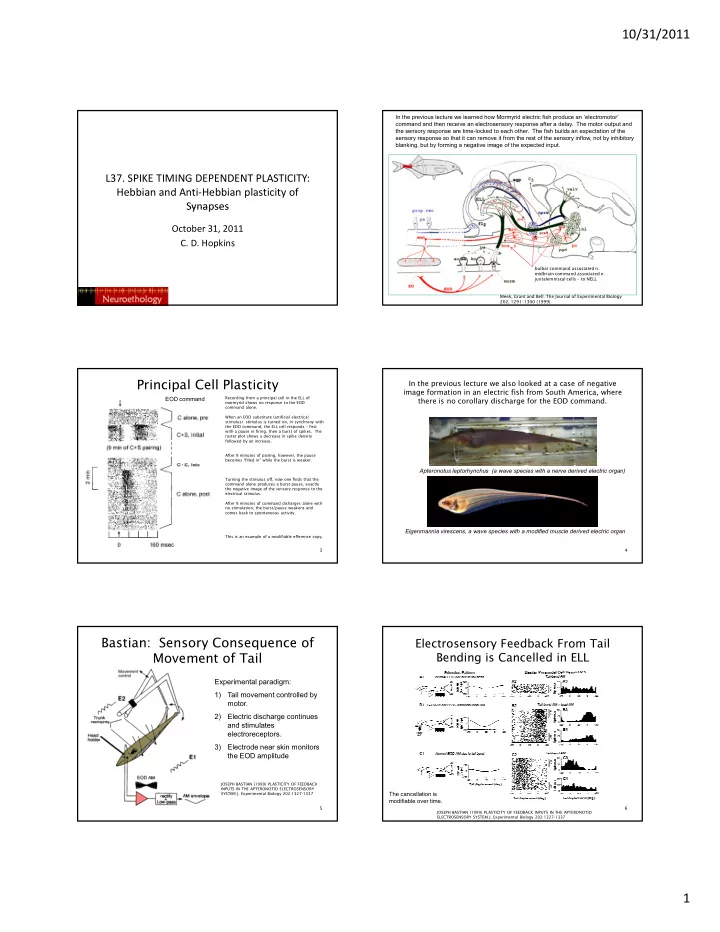
In the previous lecture we learned how Mormyrid electric fish produce an ‘electromotor’ command and then receive an electrosensory response after a delay. The motor output and the sensory response are time-locked to each other. The fish builds an expectation of the sensory response so that it can remove it from the rest of the sensory inflow, not by inhibitory blanking, but by forming a negative image of the expected input. L37. SPIKE TIMING DEPENDENT PLASTICITY: Hebbian and Anti ‐ Hebbian plasticity of Synapses Synapses October 31, 2011 C. D. Hopkins bulbar command associated n. midbrain command associated n. juxtalemniscal cells – to NELL 2 Meek, Grant and Bell. The Journal of Experimental Biology 202, 1291–1300 (1999) Principal Cell Plasticity In the previous lecture we also looked at a case of negative image formation in an electric fish from South America, where EOD command Recording from a principal cell in the ELL of there is no corollary discharge for the EOD command. mormyrid shows no response to the EOD command alone. When an EOD substitute (artificial electrical stimulus) stimulus is turned on, in synchrony with the EOD command, the ELL cell responds – first with a pause in firing, then a burst of spikes. The raster plot shows a decrease in spike density followed by an increase. After 9 minutes of pairing, however, the pause becomes “filled in” while the burst is weaker. Apteronotus leptorhynchus (a wave species with a nerve derived electric organ) Turning the stimulus off, now one finds that the command alone produces a burst-pause, exactly the negative image of the sensory response to the electrical stimulus. After 9 minutes of command disharges alone with no stimulation, the burst/pause weakens and comes back to spontaneous activity. Eigenmannia virescens, a wave species with a modified muscle derived electric organ This is an example of a modifiable efference copy. 3 4 Bastian: Sensory Consequence of Electrosensory Feedback From Tail Movement of Tail Bending is Cancelled in ELL Experimental paradigm: 1) Tail movement controlled by motor. 2) Electric discharge continues and stimulates d ti l t electroreceptors. 3) Electrode near skin monitors the EOD amplitude JOSEPH BASTIAN (1999) PLASTICITY OF FEEDBACK INPUTS IN THE APTERONOTID ELECTROSENSORY SYSTEM J. Experimental Biology 202:1327-1337 The cancellation is modifiable over time. 5 6 JOSEPH BASTIAN (1999) PLASTICITY OF FEEDBACK INPUTS IN THE APTERONOTID ELECTROSENSORY SYSTEM J. Experimental Biology 202:1327-1337 1

  2. 10/31/2011 If tail is not bent during artificial Amplitude The same experiment was done with Eigenmannia , but this time without the benefit of propioceptive input (no tail bending) modulation, there is no effect experiments were done on Eigenmannia B1) The whole body is stimulated with an amplitude modulated EOD. B2) Whole body plus a local AM B3) After 10 mintues, however the effect of the local AM goes away. B4) Whole body AM alone, now the whole body AM forms a negative image B5) after 10 minutes of AM alone,, local AM off. 7 8 Pairing of electrosensory stimulation with ventilation movements in Skate also produces a negative image of expected sensory input The principal cell in the Dorsal Octavolateral Nucleus (DON) of skate. No response to expiration, inspiration movements of gill. When ventilation is paired with electrosensory stimulus, the principal cell responds After 25 minutes, weaker response to stimulus. Now, when the stimulus goes off, the ventilation response produces a negative image 9 10 Montgomery and Bodznick, 1994 In principal cells of the mormyrid ELL, pairing a EOD The Basic Architecture command discharge with an intracellular stimulus can result in formation of negative image (only under strong stimulation) The principle cell in response to EODc + weak intracellular, no plasticity In response to strong intracellular stimulus which evokes a broad spike, plasticity occurs. Bell, C. C., Caputi, A., Grant, K. and Serrier, J. (1993). Storage 11 of a sensory pattern by anti-hebbian synaptic plasticity in an electric fish. Proc Natl Acad Sci U S A 90, 4650-4654. 1. Bell, C. C., Han, V. and Sawtell, N. B. (2008). Cerebellum-like structures and their implications for cerebellar function. Annu Rev Neurosci 31, 1-24. 2

  3. 10/31/2011 Theory to Explain Negative Image Sensory input arrives from below and stimulates pyramidal cell to fire. Predictive input comes from above through parallel fibers. Synaptic modification changes strength of predictive input as a consequence of simultaneous action. 13 14 Spike timing dependent plasticity is anti-hebbian. If the broad spike occurs Recording Plasticity in Slice after the EPSP, the synaptic weight is decreased. If the broad spike occurs before the EPSP, it strengthens. This sculpts a negative image of the Preparation expected input. Recording intracellularly from an acausal causal ELL cell from mormyromast region of mormyrid ELL. Stimulate parallel fibers with two sets of electrodes in molecular layer of ELL. T (post-pre) Percentage change in excitatory postsynaptic potential (EPSP) amplitude plotted against the delay between EPSP onset and the broad spike peak during pairing. A negative delay with regard to the previous spike and a positive delay with regard to the following spike were present for each pairing. The shorter of these two delays is 15 plotted. Filled circles, significant changes; open circles, non-significant changes (at P <0.01). animation rule Note: the sign of the time axis here is reversed compared to previous slide Bell CC, Han VZ, Sugawara Y, Grant K. 1997. Synaptic plasticity in a cerebellum-like structure depends on temporal order. Nature 387:278–81 presynaptic post synaptic spike spike before before pre post T(pre-post) Defining Spike- Timing- Dependent Plasticity (A) A presynaptic cell connected to a postsynaptic cell repeatedly spiking just before the latter is in part causing it to spike, while the opposite order is acausal. (B) In typical STDP, causal activity results in long- term potentiation (LTP), while acausal activity elicits long-term depression (LTD; Markram et al., 1997b; Bi and Poo, 1998; Zhang et al., 1998). At some cortical synapses, the temporal window for LTD (dashed gray line) is extended (Feldman, 2000; Sjöström et al., 2001). These temporal windows are often also activity dependent, with LTP being absent at low-frequency (gray continuous line, Markram et al., 1997b; Sjöström et al., 2001), and postsynaptic bursting relaxing the LTD timing requirements to hundreds of milliseconds (Debanne et al., 1994; Sjöström et al., 2003). 17 18 Henry Markram1*,Wulfram Gerstner1 and Per Jesper Sjöström2,3 Frontiers in Synaptic Neuroscience. August 2011 3

  4. 10/31/2011 normal STDP (Hebbian) reversed STDP (antiHebbian) Donald Hebb “ Let us assume that the persistence or repetition of a reverberatory activity (or “trace”) tends to induce lasting cellular changes that add to its stability.[. . .] When an axon of cell A is near enough to excite a cell B and repeatedly or persistently takes part in firing it, some growth process or metabolic change takes place in one or both cells such that A’s efficiency, as one of the cells firing B, is increased.” (Hebb, 1949). 20 Henry Markram1*,Wulfram Gerstner1 and Per Jesper Sjöström2,3 Frontiers in Synaptic Natalia Caporale and Yang Dan (2008) Neuroscience. August 2011 Cerebellum has always attracted neuroscientists interested in how the brain works. The orderly structure, the size, the individuality of the cellular Methods (continued) players, the complexity. Identification of Mossy Fibers in Egp. 1) Previous studies by Bell (1991) recorded from cells in EGp with similar properties and found * no synaptic activity, *spikes arising directly from baseline. 2) Several fills with biocytin (3 from proprioceptive input, 2 with EODc input) 3) No neurons responded to both EODc and propioception. 4) Pairing of EODCD synaptic activity and narrow spike activity by averaging responses before and after EODCD, narrow spikes removed. Ramón y Cajal 1894 Brains of vertebrates color coded by brain area. Cerebellum in orange The main players in cerebellar function are: Granule cells: which receive input from mossy fibers, and send their outputs to Purkinje cells via parallel fibers Purkinje cells: the “Principal Cell” of the Cerebellum Climbing fibers: bring “error signals” from the inferior olive to ONE Purkinje cell. 4

Download Presentation
Download Policy: The content available on the website is offered to you 'AS IS' for your personal information and use only. It cannot be commercialized, licensed, or distributed on other websites without prior consent from the author. To download a presentation, simply click this link. If you encounter any difficulties during the download process, it's possible that the publisher has removed the file from their server.

Recommend


More recommend