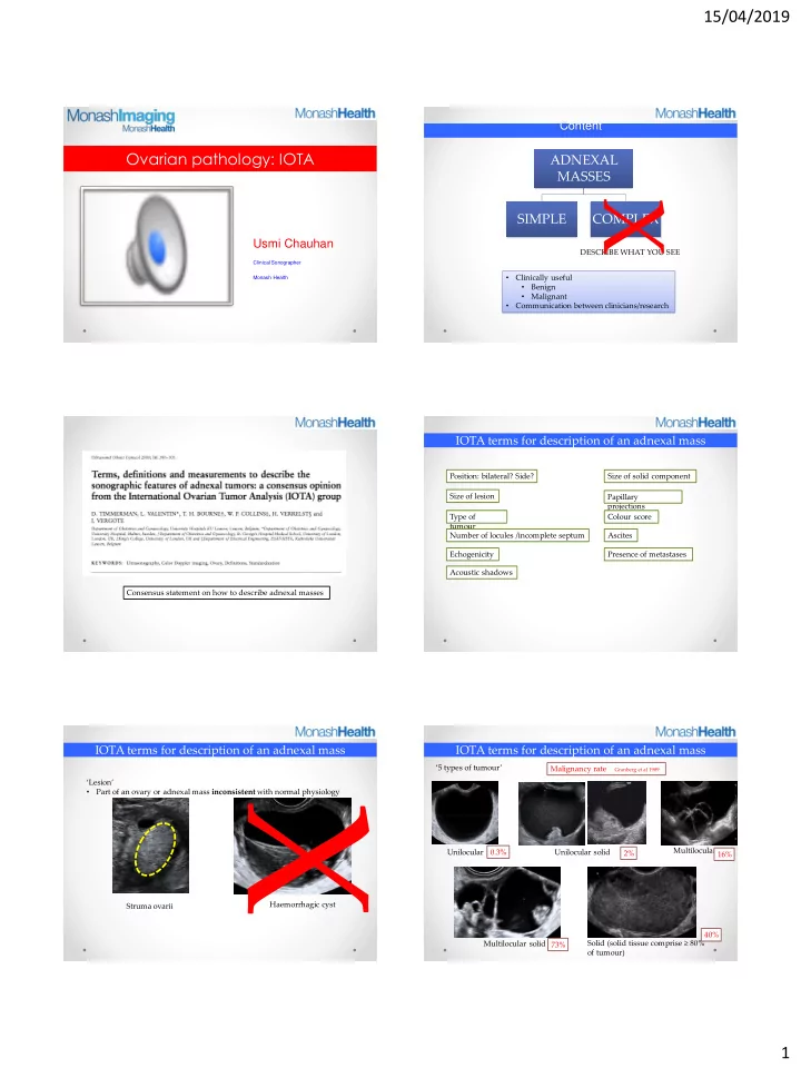
X Multilocular Unilocular 0.3% Unilocular solid 2% 16% - - PDF document

x
SMART_READER_LITE
LIVE PREVIEW

X Multilocular Unilocular 0.3% Unilocular solid 2% 16% - - PDF document

15/04/2019 Content Ovarian pathology: IOTA ADNEXAL MASSES X SIMPLE COMPLEX Usmi Chauhan DESCRIBE WHAT YOU SEE Clinical Sonographer Clinically useful Monash Health Benign Malignant Communication between


slide-1
SLIDE 1

15/04/2019 1

Ovarian pathology: IOTA

Usmi Chauhan

Clinical Sonographer Monash Health

Content

  • Clinically useful
  • Benign
  • Malignant
  • Communication between clinicians/research

DESCRIBE WHAT YOU SEE

ADNEXAL MASSES SIMPLE COMPLEX

X

Consensus statement on how to describe adnexal masses

IOTA terms for description of an adnexal mass

Position: bilateral? Side? Size of lesion Type of tumour Number of locules /incomplete septum Echogenicity Acoustic shadows Size of solid component Papillary projections Colour score Ascites Presence of metastases

IOTA terms for description of an adnexal mass

‘Lesion’

  • Part of an ovary or adnexal mass inconsistent with normal physiology

Struma ovarii Haemorrhagic cyst

X

IOTA terms for description of an adnexal mass

‘5 types of tumour’ Solid (solid tissue comprise ≥ 80%

  • f tumour)

Unilocular Unilocular solid Multilocular Multilocular solid Malignancy rate Granberg et al 1989 0.3% 2% 16% 73% 40%

slide-2
SLIDE 2

15/04/2019 2

IOTA terms for description of an adnexal mass

‘5 types of cyst contents’ Mixed Anechoic Low level Ground glass Haemorrhagic

IOTA terms for description of an adnexal mass

‘Solid component’

  • A structure that has echogenicity suggestive of tissue (ovarian stroma)
  • NOT ‘white ball’ in a dermoid cyst
  • NOT blood clot, or mucous
  • Concave
  • Probe pressure
  • Colour Doppler

IOTA terms for description of an adnexal mass

‘Papillary projection’

  • Protrusion of solid tissue into a cyst cavity ≥ 3mm (height)
  • Protrusions < 3mm (height) = irregularities
  • Papillary projections = solid component

Solid component but not PP Irregularity Papillary projection

IOTA terms for description of an adnexal mass

‘Septum’

  • Thin strand of tissue that runs from one internal cyst surface to another

Incomplete septum

  • does not reach the opposite wall of the cystic structure in some scanning

planes (seen in diseased tubes) Septum Incomplete septum

IOTA terms for description of an adnexal mass

‘Shadowing’

IOTA terms for description of an adnexal mass

‘Ascites’

  • Fluid outside pouch of Douglas
slide-3
SLIDE 3

15/04/2019 3

IOTA terms for description of an adnexal mass

‘IOTA colour score’

  • Adjust settings: maximize detection of flow without artifacts (3-6cm/s; PRF 0.3-0.6 kHz)

Score 1 (none) Score 2 (minimal) Score 3 (moderate) Score 4 (strong)

How do we use IOTA: practical applications

IOTA Easy descriptors Pattern recognition Simple rules ADNEX LR 1/LR 2

IOTA: Easy descriptors

Unilocular cyst, ground glass echogenicity, +/- wall nodularity. Premenopausal woman ENDOMETRIOMA Unilocular cyst, mixed echogenicity (white ball, dot-dash), shadowing, Premenopausal DERMOID CYST Unilocular cyst, anechoic cyst fluid, regular walls,< 10 cm SIMPLE CYST OR CYSTADENOMA All other unilocular cysts with regular walls BENIGN Mass > 50 years CA125 > 100 IU/mL MALIGNANT Mass At least moderate blood flow Postmenopausal Ascites MALIGNANT

IOTA: Easy descriptors

SIMPLE CYST OR CYSTADENOMA

  • Unilocular cyst
  • Anechoic cyst fluid
  • Regular walls
  • < 10 cm

IOTA: Pattern recognition

ENDOMETRIOMA Wall nodularity (blood clot/fibrin) (Not papillary projection)

  • Unilocular cyst
  • Ground glass

echogenicity

  • +/- wall nodularity
  • premenopausal woman

IOTA: Pattern recognition

DERMOID CYST

  • Unilocular cyst
  • Mixed echogenicity

(white ball, dot-dash)

  • Shadowing
  • Premenopausal
slide-4
SLIDE 4

15/04/2019 4

IOTA: Pattern recognition

HAEMORRHAGIC CYST Reticular pattern Retracting clot

IOTA: Pattern recognition

MALIGNANT

  • Mass with at least moderate blood flow
  • Postmenopausal
  • Ascites

MALIGNANT

  • Mass
  • > 50 years
  • CA125 > 100 IU/mL

And/or

IOTA: Pattern recognition

HYDRO (PYO/HEMATO) SALPINX

Serpiginous Incomplete septae Cog wheeling Beads on a string

PERITONEAL PSEUDOCYST

IOTA: Pattern recognition

  • Conforms to shape of peritoneal cavity
  • Prior pelvic surgery

Ovary suspended in edge of cyst

Solid Pelvic Tumours

  • Myoma (uterine)
  • Fibroma
  • Thecoma
  • Brenner tumour
  • Leydig cell tumour
  • Malignancies

Peduculated fibroid Fibroma

  • Round,
  • val

lobulated;

  • Stripy

shadows

  • Min

vascularity

  • May mimic pedunculated fibroid

Broad ligament fibroid Ovarian malignancy

Irregular contour Irregular echogenicity Increased vascularity

Dysgerminoma + yolk sac tumour

Solid Pelvic Tumours

Neuroendocrine tumour of ovary Granulosa cell tumour

slide-5
SLIDE 5

15/04/2019 5

IOTA: Simple Rules

Timmermen et al UOG 2008

IOTA: Simple Rules

B-rules M-rules Unilocular cysts. Irregular solid tumour. Presence of solid component where the largest solid component <7 mm. Ascites. Acoustic shadowing. At least four papillary structures. Smooth multi-locular tumour with a largest diameter <10 cm. Irregular multilocular solid tumour with largest diameter ≥ 10 cm. No blood flow. Very strong blood flow.

IOTA: ADNEX model

https://www.iotagroup.org/sites/default/files/adnexmodel/IOTA%20-%20ADNEX%20model.html

CONCLUSION

  • 1. Describe the mass instead of using the term ‘complex’
  • 2. Be familiar with easy descriptors and pattern recognition
  • 3. If uncertain, use Simple Rules +/- ADNEX

THANK YOU

THANK YOU