
SLIDE 1
Meng Cui HHMI Janelia Farm Research Campus
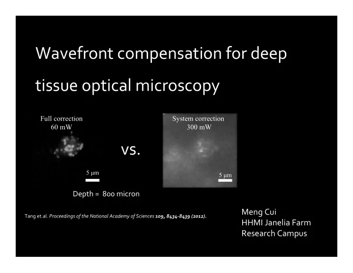
Wavefront compensation for deep tissue optical microscopy
5 μm
Full correction 60 mW
5 μm
System correction 300 mW
vs.
Depth = 800 micron
Tang et.al. Proceedings of the National Academy of Sciences 109, 8434‐8439 (2012).