
SLIDE 1
ANTIGEN CATEGORIZATION
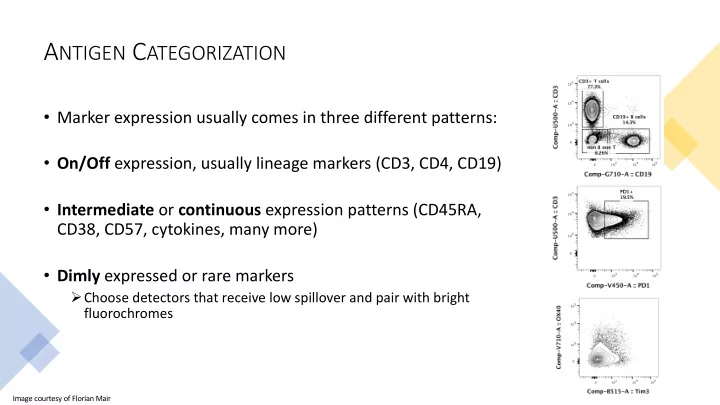
- Marker expression usually comes in three different patterns:
- On/Off expression, usually lineage markers (CD3, CD4, CD19)
- Intermediate or continuous expression patterns (CD45RA,
CD38, CD57, cytokines, many more)
- Dimly expressed or rare markers
ØChoose detectors that receive low spillover and pair with bright fluorochromes
Image courtesy of Florian Mair