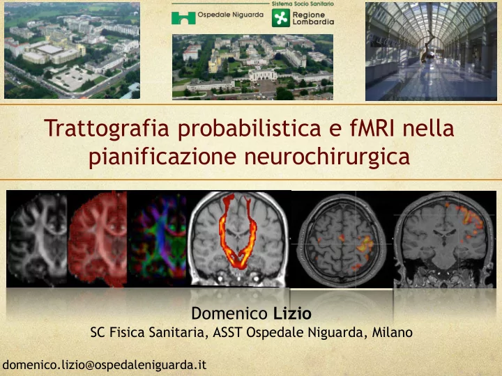
SLIDE 1 Trattografia probabilistica e fMRI nella pianificazione neurochirurgica
Domenico Lizio
SC Fisica Sanitaria, ASST Ospedale Niguarda, Milano
domenico.lizio@ospedaleniguarda.it
SLIDE 2 Basic Anatomy
The brain is full of
- neurons. These are
- rganised into two
types of “tissues”:
- Grey Matter (GM)
- White Matter (WM)
MRI Post-Mortem Grey Matter White Matter
SLIDE 3
Neurons are densely connected and have many dendrites Axons conduct electrical signals and are surrounded by myelin Myelin is a major factor in determining the MR signal and contrast
Basic Anatomy
SLIDE 4 Applications
Neuroimaging
fMRI: finger-tapp. mano dx
Functional MRI
Mean diffusivity (MD) Fractional anisotropy (FA) Principal diffusion direction (PDD)
Diffusion MRI Structural MRI
T1-weighted T2-weighted Proton Density
SLIDE 5 Structural MRI
- Study of taxi drivers showing structural plasticity
Applications of MRI in Neuroimaging
Maguire et al.,PNAS, 2000
SLIDE 6
Diffusion MRI
- White matter integrity - imaging tissue nature change
- Differences in brain connectivity in multiple tracts
Green = white matter tract ; Red/Yellow = statistically significant change in FA
Roine et al., Molecular Autism, 2015
SLIDE 7
Menke et al., Brain 2013
Look at tracts connected to regions
Diffusion MRI Structural MRI
SLIDE 8
Johansen-Berg, et al., Brain 2002
Functional MRI (Task)
Group: Correlations with improvement Single subject: responder
SLIDE 9 Functional MRI (Resting)
- Altered functional connectivity
SLIDE 10
Diffusion MRI Functional MRI (Task)
Bartsch et al., JMRI 2006
Structural MRI
SLIDE 11 Functional MRI
- Measures haemodynamic response to neural activity
- Task-based or resting-state-connectivity
- Intrinsic contrast BOLD (Blood Oxygen Level Dependent)
- Take many fast images (EPI): 5-30 min scan
SLIDE 12 Magnetic field perturbed Dephasing of nearby spins Loss of signal
BOLD Effect
Diamagnetic (same as tissue)
Paramagnetic Δχ≈0.2 ppm
SLIDE 13
Magnetic field less perturbed Less dephasing More signal
BOLD Effect
Increased Neuronal Activity
CMRO2 CBV CBf
SLIDE 14
Analysis of Functional MRI
Task FMRI Resting-State fMRI & Connectivity
SLIDE 15 Images - Low Resolution FMRI
- A sequence of low resolution T2*-weighted volumes are
taken during the FMRI experiment
- Optimised for BOLD sensitivity and speed
- Take one volume every 1-3 seconds
- Often take around 200 volumes (10 minutes)
- An fMRI volume is shown here in orthogonal view
SLIDE 16 Functional MRI Artefacts
Distortion Signal Loss Physiological Noise
Hardware-related Physiological
... plus most diffusion artefacts (not eddy currents) and all structural artefacts
- Distortion due to B0 inhomogeneity (air in sinuses) -
acquisition-and-analysis related fixes needed (fieldmap)
- Physiological noise is more problematic near the brainstem -
acquire physiological measurements & do analysis fix
- Motion can also be a significant problem (some analysis fixes)
SLIDE 17 Functional MRI ‘‘Limitations’’
- Does not measure electrical activity
- Does not measure metabolic activity
- BOLD-FMRI is qualitative
- Sensitive to fast imaging artefacts
- Need good T2* sensitivity
- causes lost signal in inferior regions
SLIDE 18 Functional MRI Acquisition
Basic tips for acquisition
- Use optimised sequences/protocol for your scanner/site
- Get fieldmap (B0) for compensating distortion/signal-loss -
blip-up-blip-down is not an option for functional MRI
- For inferior-frontal/temporal areas apply acquisition
techniques to minimise signal loss - e.g. thin slices, slice angulation, z-shims, parallel imaging, ...
- Isotropic voxels (or close) are better for analysis generally
- For small FOV also take one single whole-brain EPI
- Biggest interaction of exp. design-acquisition-analysis so
think carefully about all parts before acquiring data!
SLIDE 19 Diffusion MRI
- Measures microstructure directionality and “integrity”,
particularly in WM
- Provides information on anatomical connectivity
- Need to acquire many “directions”: 5-30 min scan
SLIDE 20
- Random motion of particles due to thermal energy
- Water molecules collide and experience net displacement
- Displacement described by diffusion coefficient (D)
- Normally, diffusion is isotropic (equal in all directions)
What is diffusion?
SLIDE 21
- Diffusion is restricted by tissue boundaries, membranes, etc
- Marker for tissue microstructure (healthy and pathology)
Why is diffusion interesting?
SLIDE 22
Water can diffuse more freely along white matter fibres than across them
Diffusion anisotropy in white matter
SLIDE 23
Diffusion anisotropy in white matter
Diffusion in white matter fibres is “anisotropic” Directionality of diffusion tells us about fibre integrity/structure and orientation
SLIDE 24
Displacement due to diffusion is approximately ellipsoidal Eigenvectors = axes of ellipsoid (direction of fibres) Eigenvalues = size of axes (strength of diffusion)
The diffusion tensor
SLIDE 25
Principal diffusion direction (PDD): what direction is greatest diffusion along? Info about fibre orientation Fractional anisotropy (FA): how elongated is the ellipsoid? Info about fibre integrity The diffusion tensor: Useful quantities Mean diffusivity (MD) Info about tissue integrity
The diffusion tensor: Useful quantities
SLIDE 26
Mean diffusivity (MD) Fractional anisotropy (FA) Principal diffusion direction (PDD)
Diffusion tensor imaging
At each voxel, fit the diffusion tensor model Can then calculate MD, FA, PDD from fitted parameters
SLIDE 27
Mean diffusivity (MD)
Control MD Acute Stroke
Mean diffusion coefficient across all directions Correlate of tissue integrity (white and gray matter) Example: MD is altered in acute and chronic stroke
SLIDE 28
Inequality of diffusion coefficient across different directions High in regions where diffusion is most directional Relates to integrity of white matter fibre bundles
Fractional Anisotropy (FA)
SLIDE 29
Principal diffusion direction (PDD)
Direction along which greatest diffusion occurs Relates to direction of fibre orientations Typically, will use this as starting point for fibre tracking
SLIDE 30
Diffusion tractography
Follow PDD to trace white matter fibers (“tractography”)
SLIDE 31
More complicated models: Crossing fiber populations
SLIDE 32 Diffusion tractography: Determionistic vs Probabilistic
- Deterministic assumes a single orientation at each voxel: one
streamline per seed voxel.
- Probabilistic assumes a distribution of orientations: multiple streamline
samples per seed voxel (drawn from probability distribution)
SLIDE 33
If diffusion is present, gradients cause a drop in signal. Greater Diffusion = Less Signal
Acquiring the image
SLIDE 34 Diffusion MRI Artefacts
Eddy Currents Hardware-related
+ Bulk/Pulsatile Motion … plus all the structural artefacts
- Eddy currents: both acquisition and analysis fixes available
- Distortion due to B0 inhomogeneity (air in sinuses)- acquisition-
and-analysis related fixes needed
- Bulk motion is corrected for in acquisition (navigators)
- Pulsatile motion is more problematic
SLIDE 35
Diffusion gradients encode tiny displacement Subject motion is also accidentally encoded Image artefacts if we try to combine data from multiple excitations (different motion)
Motion in Diffusion MRI
Linescan diffusion image
SLIDE 36
Can motion be avoided?
Subject restraints can reduce bulk motion, but... ...in the brain, there is significant non-rigid motion from cardiac pulsatility cardiac gating helps, but brain is never very still!
SLIDE 37
Single-shot echo-planar imaging (EPI)
Single-shot imaging freezes motion Most common method is echo-planar imaging (EPI) Images have serious distortion and limited resolution
SLIDE 38
Eddy Currents
Diffusion gradients create large eddy currents, which persist into acquisition window Distort the k-space trajectory, casing shears/scaling of images Eddy currents “resist” gradient field changes effective gradient fields
SLIDE 39 Diffusion MRI “Limitations”
- Does not measure axon size/density directly
- Does not measure single fibres (only average groups)
- More difficult to deal with crossing/kissing fibres
- Quantitative local measurements, but not
connectivity
- More difficult to do in pulsatile regions (e.g.
brainstem)
- More restricted by hardware and SNR
- Sensitive to fast imaging artefacts
SLIDE 40 Diffusion MRI: Acquisition
Basic tips for acquisition
- Best parameters can be quite hardware dependent (esp.
gradients) so check what is optimised for your scanner
- In general, b-value of 1000-1500 s/mm2 and 60+ directions
(tractography) or 12+ directions (FA, etc.) tend to give good results (but the more directions the better)
- Get one b=0 image for every 8-10 diffusion-weighted images
- Get a fieldmap (B0) for distortion correction - or alternatively, a
blip-up-blip-down
- Isotropic voxels (or close) are better for analysis generally
- Do not do oversampling on scanner (sometimes the default)
- Both single/multi-shell give good tractography
SLIDE 41
Parameter Value Relevant points
TE (Echo Time) 100 ms Limited by b-value Matrix size /Resolution 128x128 / 2 mm Limited by distortion, SNR Number of directions 6-60 Lower limit: tensor model Upper limit: scan stime b-value 1000 s/mm2 Larger b = more contrast Smaller b = more signal
Typical Diffusion Imaging Parameters
SLIDE 42
Diffusion MRI: Analysis
Basic stages in the diffusion analysis pipeline: Eddy Current & Motion Correction Fibre/ direction modelling Probalistic Tractography ‘‘Tensor’’ Fitting e.g. FA, MD
SLIDE 43 Behrens T .E.J. et al. NeuroImage, 2006 www.fmrib.ox.ac.uk/fsl
FSL tractography pipeline
SLIDE 44
Case Studies
SLIDE 45 Clinic: patient suffering from focal epilepsy with multiple daily motor seizures prevalent in the right upper limb. Diagnosis: clearly visible focal cortical dysplasia with FLAIR sequences contiguous to the primary motor region of the hand in the ascending frontal convolution. --> SEEG Pre-surgical question: CST and ThC sn fibre reconstruction in the hand component..
Case Study No. 1: Motor-sensitive system
FLAIR T1W-3D SEEG
SLIDE 46 P6 - CST mano (ST+MEP) I2 - CST piede (ST+MEP) P1 - ThC mano (SEP) Q5 - ThC piede (SEP)
SEED CST: peduncle cerebral sx
(identified by DTI colormap)
SEED ThC: nVPL sx
(identified with connectivity- based seed classification)
WAYPOINTs: M1 - S1 hand (GM), exact points of the CST and ThC hand beams (WM) WAYPOINTS functional by SEEG information
Case Study No. 1: Motor-sensitive system
SLIDE 47
CST hand (RED) - CST foot (ORANGE) ThC hand (BLUE) - ThC foot (BLUE)
Case Study No. 1: Motor-sensitive system
SLIDE 48 Clinic: patient suffering from partial epilepsy with multiple daily motor seizures with prevalent involvement of the right shoulder. Diagnosis: clearly visible focal cortical dysplasia with FLAIR sequences contiguous to the primary motor region of the hand in the ascending frontal convolution. Pre-surgical question: identification of cortical areas and corresponding fibres responsible for the primary motor and sensitive functions of the contralateral hand.
T1W-3D FLAIR
Case Study No. 2: Motor-sensitive system
SLIDE 49 fMRI: finger-tapp. Right hand SEED CST: peduncle Cerebral sx
(identified by DTI colormap)
WAYPOINTs SEED ThC: nVPL sn
(identified with connectivity- based seed classification)
Case Study No. 2: Motor-sensitive system
SLIDE 50
SEEG planning (subsequent to FT) Case Study No. 2: Motor-sensitive system
SLIDE 51 Clinic: patient suffering from partial focal epilepsy with suspected left temporo-occipital ZE. Diagnostics: Negative MRI. --> SEEG Pre-surgical question: fibre tracking Radiation Optics sx.
Case Study No. 3: Visual System
FLAIR SEEG
SLIDE 52 V4 - OR sup (ST+VEP) V4 - OR sup (ST+VEP) V4 - OR sup (ST+VEP) F8-9 - OR sup (wm VEP)
WAYPOINTs OR sup:
- cont. V4 (gm)
- cont. F8-9 (wm)
SEED OR:
lGNB sx
O2 - OR inf (ST+VEP) O2 - OR inf (ST+VEP) O2 - OR inf (ST+VEP) E8 - OR inf (wm VEP)
WAYPOINTs OR inf:
- cont. O2 (gm)
- cont. E8 (wm)
WAYPOINTs: V1 over and undercalcium (GM), certain points along OR (WM)
Case Study No. 3: Visual System
SLIDE 53
SLIDE 54
SLIDE 55
Case Study No. 3: Visual System
SLIDE 56
Injury in the Seed region used for the reconstruction of the CST fasciculus.
Case Study No. 4:
Patient with Meningioma of the Tentory in the region of the cerebellar pedicle in the study for SRS with GK
SLIDE 57 Co-registered fasciculus with AX_Helmet
- Red-Yellow: CST Hand
- Blue-Blue: CST Foot
(overlap near the lesion)
SLIDE 58 Elaborazione per Trattamento in GK
Plan Plan1 Target Meningioma Total Treatment Time [min] 52.0 Prescription Isodose [%] 50 Prescription dose [Gy] 6.5 Maximum dose [Gy] 13.0 Average dose[Gy] 8.9 Minimum dose [Gy] 5.4 Number of shots 38(1) Gamma angle 90 TV - Target Volume [cm³] 3.884 Intersection TVÇPIV [cm³] 3.754 PIV50 - 50% Prescription Isodose Volume [cm³] corresponding to half of Prescription Dose 11.568 PIV - Volume [cm³] of the Prescription Isodose 4.185 Conformity Index - TVÇPIV/TV 0.97 Selectivity index - TVÇPIV/PIV 0.90 Gradient index - PIVh/PIV 2.76 Paddick Index: Conformity x Selectivity 0.867
SLIDE 59
Processing for GK treatment
SLIDE 60
SLIDE 61
SLIDE 62
SLIDE 63
- The diffusion tensor imaging (DTI) enables to investigate
cerebral white matter integrity non-invasively.
- The use of the probabilistic tractography method and
functional magnetic resonance imaging for the planning of the pre-surgical study is very useful. The main limitations of FT-DTI are:
- 1. reliability of the reconstructions following the brain-
shift
- 2. probability threshold to be used.
The main limitations of fMRI are: Design of experiment Requires adequate task performance Accurate and reproducible fMRI strongly depends on the patient’s cooperation
Conclusion
SLIDE 64 Acknowledgements
Team
- Dr. Luca Berta
- Dr. Paola Colombo
- Dr. Marco Felisi
- Dr. Stefania Nici
- Dr. Alberto Torresin
SLIDE 65