BOLD fMRI and fcMRI fcMRI in the Pediatric in the Pediatric BOLD - - PowerPoint PPT Presentation

bold fmri and fcmri fcmri in the pediatric in the
SMART_READER_LITE
LIVE PREVIEW

BOLD fMRI and fcMRI fcMRI in the Pediatric in the Pediatric BOLD - - PowerPoint PPT Presentation

BOLD fMRI and fcMRI fcMRI in the Pediatric in the Pediatric BOLD fMRI and Brachial Plexus Injury Population Population Brachial Plexus Injury Jacques A. Machol IV, MD, 1,2 Rupeng Li, MD, PhD, 2 Nicholas A. Flugstad,MD, 1,2 Ji-Geng Yan, MD,


slide-1
SLIDE 1

BOLD fMRI and BOLD fMRI and fcMRI fcMRI in the Pediatric in the Pediatric Brachial Plexus Injury Brachial Plexus Injury Population Population

Jacques A. Machol IV, MD,1,2 Rupeng Li, MD, PhD,2

Nicholas A. Flugstad,MD,1,2 Ji-Geng Yan, MD, PhD,1 James S. Hyde, PhD,2 Hani S. Matloub, MD1 Medical College of Wisconsin, Departments of Plastic Surgery1 & Biophysics2, Milwaukee, WI, USA No Financial Disclosures

Introduction

  • Brachial plexus birth palsy (BPBP) occurs in

approximately 1/1000 live births1,2

  • The most common of these palsies involve the

C5-C6 roots of the brachial plexus3

  • A number of surgical procedures for

reconstruction have been introduced4

  • Likewise, blood oxygen level dependent (BOLD)

Functional MRI (fMRI) has provided a reliable method for indirectly studying task-induced cerebral neuronal activity5-6

  • This imaging exploits changes in deoxygenated

hemoglobin (dHb) concentrations, which, in turn, act as an endogenous paramagnetic contrast agent

Introduction

Image: Astolfi et al. IJBEM. Vol. 6, No. 2. 2004

Figure 1. Signal in fMRI

slide-2
SLIDE 2

Introduction

  • Cortical metabolism is almost exclusively aerobic
  • Thus, the local dHb to Hb ratio measured by fMRI

can be interpreted as an indirect measurement of neuronal activity

  • Likewise, Functional Connectivity MRI (fcMRI)

uses spontaneous low frequency BOLD fluctuations to demonstrate cortical connectivity7-9

  • Our laboratory has extensive experience utilizing

fMRI to reveal cortical plasticity following peripheral nerve injury and repair10-15

  • No human studies assessing cortical changes

after BPBP exist

Introduction

BOLD fMRI and BOLD fMRI and fcMRI fcMRI in the Pediatric in the Pediatric Brachial Plexus Injury Brachial Plexus Injury Population Population

Jacques A. Machol IV, MD,1,2 Rupeng Li, MD, PhD,2

Nicholas A. Flugstad,MD,1,2 Ji-Geng Yan, MD, PhD,1 James S. Hyde, PhD,2 Hani S. Matloub, MD1 Medical College of Wisconsin, Departments of Plastic Surgery1 & Biophysics2, Milwaukee, WI, USA

1

  • 1

Figure 1. 9.4T fMRI during C7 (top) and median nerve stimulation (bottom)10

Li R, Machol IV JA, et al. Muscle Nerve. 2013

slide-3
SLIDE 3

Introduction

  • We employ 3T BOLD fMRI and fcMRI in a pre-
  • perative pediatric BPBP patient and a healthy

adult

  • Assess post-injury cortical changes using Air-

Puffer somatosensory stimulation16

  • Post central gyrus chosen as the region of

principal evaluation (primary sensory cortex)17

  • fMRI and fcMRI of the BPBP patient’s injury side

sensory cortex is contrasted to: – Non-injury side (internal control) – Healthy adult cortex

  • We hypothesize that there will be significant

differences in BOLD signal noted for both comparisons

Introduction

BOLD fMRI and BOLD fMRI and fcMRI fcMRI in the Pediatric in the Pediatric Brachial Plexus Injury Brachial Plexus Injury Population Population

Jacques A. Machol IV, MD,1,2 Rupeng Li, MD, PhD,2

Nicholas A. Flugstad,MD,1,2 Ji-Geng Yan, MD, PhD,1 James S. Hyde, PhD,2 Hani S. Matloub, MD1 Medical College of Wisconsin, Departments of Plastic Surgery1 & Biophysics2, Milwaukee, WI, USA

Figure 2. Human post central gyrus (red). Primary sensory cortex.

Image: http://commons.wikimedia.org/wiki/File:Postcentral_gyrus_3d.png

slide-4
SLIDE 4

Methods

  • 10 mo Female
  • Left C5-C6 BPBP
  • 0/5 Ext. Rotators
  • 0/5 Post. Deltoid
  • No withdrawal with pinch of lateral deltoid
  • Modified Mallet Classification

– Global Abduction III – External Rotation III – Hand to Neck I – Hand to Spine II – Hand to Mouth I

Methods

BOLD fMRI and BOLD fMRI and fcMRI fcMRI in the Pediatric in the Pediatric Brachial Plexus Injury Brachial Plexus Injury Population Population

Jacques A. Machol IV, MD,1,2 Rupeng Li, MD, PhD,2

Nicholas A. Flugstad,MD,1,2 Ji-Geng Yan, MD, PhD,1 James S. Hyde, PhD,2 Hani S. Matloub, MD1 Medical College of Wisconsin, Departments of Plastic Surgery1 & Biophysics2, Milwaukee, WI, USA

Table 1. Upper Extremity Functional Exam using the Medical Research Council (MRC) Scale for Muscle Strength. 0: no function – 5: contracts against full resistance. Testing was performed within the best ability given the patient’s age.

slide-5
SLIDE 5

Methods

  • Modified Mallet Classification

– Global Abduction III – External Rotation III – Hand to Neck I – Hand to Spine II – Hand to Mouth I

  • The C5-C6 pathology was verified with pre-
  • perative EMG
  • Post-scan surgical exploration and intra-
  • perative EMG confirmed neuroma at C5-C6

(during nerve transfer)

Methods

BOLD fMRI and BOLD fMRI and fcMRI fcMRI in the Pediatric in the Pediatric Brachial Plexus Injury Brachial Plexus Injury Population Population

Jacques A. Machol IV, MD,1,2 Rupeng Li, MD, PhD,2

Nicholas A. Flugstad,MD,1,2 Ji-Geng Yan, MD, PhD,1 James S. Hyde, PhD,2 Hani S. Matloub, MD1 Medical College of Wisconsin, Departments of Plastic Surgery1 & Biophysics2, Milwaukee, WI, USA

Figure 3. Intra-operative image of nerve transfer after identification of C5-C6 neuroma. Thoracodorsal n. to Axillary n. (side to side) with neurolysis was completed.

slide-6
SLIDE 6

Methods

  • Children’s Hospital of Wisconsin IRB and MCW

MRI Safety approval obtained

  • GE 3.0T short-bore utilized for MRI scans
  • A timed air-puff stimulator using CO2 gas was

connected to two tubes to intra-MRI arm cradles

  • One tube was designated the RUE and the other

was directed to the LUE

  • The lateral deltoid was selected for stimulation
  • C5-C6 dermatome

Methods

BOLD fMRI and BOLD fMRI and fcMRI fcMRI in the Pediatric in the Pediatric Brachial Plexus Injury Brachial Plexus Injury Population Population

Jacques A. Machol IV, MD,1,2 Rupeng Li, MD, PhD,2

Nicholas A. Flugstad,MD,1,2 Ji-Geng Yan, MD, PhD,1 James S. Hyde, PhD,2 Hani S. Matloub, MD1 Medical College of Wisconsin, Departments of Plastic Surgery1 & Biophysics2, Milwaukee, WI, USA

Figure 4. Air-Puffer Mechanism. AIRSTIM™ controlled L and R UE tubes to bilateral, intra- scanner, custom machined, G-10 fiberglass arm

  • cradles. Each arm cradle was padded prior to
  • use. This design prevented arm flexion and

allow specific dermatome sensory targeting (C5

  • C6). CO2 gas was regulated to 60psi.

.

slide-7
SLIDE 7

Methods

  • Pre-op 3T BOLD fMRI imaging was performed

(BPBP patient)

  • Air-puff stimulus to the left (injury) and the right

(non-injury) sides during the EPI phase - completed in duplicate during separate imaging runs

  • fMRI of the pediatric injury side cortex was

compared to the non-injury side cortex

  • The injury patient’s post-central gyrus cortical

function was then compared to a healthy 31 year old adult using identical somatosensory stimulus BOLD fMRI protocols

  • fcMRI was then performed to evaluate sensory

connectivity differences between the healthy adult and the BPBP patient

Methods

  • Echo Planar Image (EPI) data from each scan

was averaged and masked using Analysis of Functional Neuro Images (AFNI) software19

  • P-value threshold of ≤ 0.005 was set to

determine significant Voxel activation (BOLD Signal) - Voxel – Represents 2.5 mm3 (Similar to a 3D pixel)

BOLD fMRI and BOLD fMRI and fcMRI fcMRI in the Pediatric in the Pediatric Brachial Plexus Injury Brachial Plexus Injury Population Population

Jacques A. Machol IV, MD,1,2 Rupeng Li, MD, PhD,2

Nicholas A. Flugstad,MD,1,2 Ji-Geng Yan, MD, PhD,1 James S. Hyde, PhD,2 Hani S. Matloub, MD1 Medical College of Wisconsin, Departments of Plastic Surgery1 & Biophysics2, Milwaukee, WI, USA

Figure 5. Air-Puffer Stimulus Timing during the EPI phase of the BOLD fMRI. The puffer remained off for 40 seconds, then on for 20 seconds. This was repeated five times followed by a rest period of 40 seconds. 60psi of CO2 was used as stimulus in the C5-C6 dermatome. (s = seconds)

slide-8
SLIDE 8
  • Results

BOLD fMRI and BOLD fMRI and fcMRI fcMRI in the Pediatric in the Pediatric Brachial Plexus Injury Brachial Plexus Injury Population Population

Jacques A. Machol IV, MD,1,2 Rupeng Li, MD, PhD,2

Nicholas A. Flugstad,MD,1,2 Ji-Geng Yan, MD, PhD,1 James S. Hyde, PhD,2 Hani S. Matloub, MD1 Medical College of Wisconsin, Departments of Plastic Surgery1 & Biophysics2, Milwaukee, WI, USA

  • Right deltoid (non-

injury) somatosensory stimulus – BOLD Signal noted in the patient’s left post-central gyrus

Figure 1. 10 Month Female BPBP. Coronal (above) and Axial (below) fMRI during healthy right deltoid air-puff stimulation. BOLD signal noted in left post central gyrus. (crosshairs and arrow denote signal)

1

  • 1

R L +Injury

slide-9
SLIDE 9
  • Results

BOLD fMRI and BOLD fMRI and fcMRI fcMRI in the Pediatric in the Pediatric Brachial Plexus Injury Brachial Plexus Injury Population Population

Jacques A. Machol IV, MD,1,2 Rupeng Li, MD, PhD,2

Nicholas A. Flugstad,MD,1,2 Ji-Geng Yan, MD, PhD,1 James S. Hyde, PhD,2 Hani S. Matloub, MD1 Medical College of Wisconsin, Departments of Plastic Surgery1 & Biophysics2, Milwaukee, WI, USA

  • Left deltoid (injury)

somatosensory stimulus

  • Lack of BOLD signal in the post

central gyrus in the right cortex

  • Intra-cortical changes noted as

compared to the non-injury cortex

Figure 2. 10 Month Female BPBP. Coronal (above) and Axial (below) fMRI during injury left deltoid air-puff stimulation. No BOLD signal noted in right post central gyrus. (crosshairs and arrow denote signal)

1

  • 1

R L +Injury

slide-10
SLIDE 10

BOLD fMRI and BOLD fMRI and fcMRI fcMRI in the Pediatric in the Pediatric Brachial Plexus Injury Brachial Plexus Injury Population Population

Jacques A. Machol IV, MD,1,2 Rupeng Li, MD, PhD,2

Nicholas A. Flugstad,MD,1,2 Ji-Geng Yan, MD, PhD,1 James S. Hyde, PhD,2 Hani S. Matloub, MD1 Medical College of Wisconsin, Departments of Plastic Surgery1 & Biophysics2, Milwaukee, WI, USA

  • Results

1

  • 1

Healthy Adult R Side Healthy Ped. R Side Injured Ped. L Side

  • Intra-cortical variance was also

illustrated when compared to a healthy adult subject

  • The BOLD signals during the healthy limb

studies appeared to closely match (top and bottom left)

  • Whereas, the somatosensory cortical

representation of the pediatric injury side did not demonstrate BOLD signal at this significance, P < 0.005 (top and bottom right)

  • Figure 3. BOLD fMRI of Axial Healthy Adult

Left Deltoid stimulation vs. Healthy Pediatric side vs. Injury Pediatric Side

slide-11
SLIDE 11

BOLD fMRI and BOLD fMRI and fcMRI fcMRI in the Pediatric in the Pediatric Brachial Plexus Injury Brachial Plexus Injury Population Population

Jacques A. Machol IV, MD,1,2 Rupeng Li, MD, PhD,2

Nicholas A. Flugstad,MD,1,2 Ji-Geng Yan, MD, PhD,1 James S. Hyde, PhD,2 Hani S. Matloub, MD1 Medical College of Wisconsin, Departments of Plastic Surgery1 & Biophysics2, Milwaukee, WI, USA

1

  • 1

Figure 3. fcMRI of Axial Healthy Adult (above) with symmetric connectivity. Pediatric Left BPBP (below) with less network connectivity in the right cortex.

  • fcMRI demonstrated connectivity difference

between the healthy subject and the BPBP patient

  • Healthy Adult: symmetrical, bilateral

somatosensory networks are demonstrated using fcMRI techniques (Top)

  • BPBP Patient: a similar sensory network is

shown when the fcMRI seed was chosen from the healthy cortical side (L cortex for R deltoid) However, the injury side cortical network has less

  • rganization (Arrow, R cortex for L deltoid)
slide-12
SLIDE 12

Conclusions

  • This novel application of 3T BOLD fMRI

and fcMRI has demonstrated intra-cortical somatosensory functional and connectivity differences in a high BPBP patient

  • The model proposed is applicable to

demonstrate cortical sensory changes in the pre and post-operative patient with BP injuries

Conclusions

BOLD fMRI and BOLD fMRI and fcMRI fcMRI in the Pediatric in the Pediatric Brachial Plexus Injury Brachial Plexus Injury Population Population

Jacques A. Machol IV, MD,1,2 Rupeng Li, MD, PhD,2

Nicholas A. Flugstad,MD,1,2 Ji-Geng Yan, MD, PhD,1 James S. Hyde, PhD,2 Hani S. Matloub, MD1 Medical College of Wisconsin, Departments of Plastic Surgery1 & Biophysics2, Milwaukee, WI, USA

Image: Netter Atlas of Human Anatomy

slide-13
SLIDE 13

Conclusions

  • Limitations: small patient sample size, comparison to adult, and no motor or post-operative

imaging

  • Represents the early phase of prospective pre and post-operative fMRI studies
  • Goals: Evaluate cortical plasticity after nerve transfer surgery for BP injury in

the pediatric and adult populations

  • Track treatment progress or assess candidacy for nerve transfer or other

resconstructive procedures

BOLD fMRI and BOLD fMRI and fcMRI fcMRI in the Pediatric in the Pediatric Brachial Plexus Injury Brachial Plexus Injury Population Population

Jacques A. Machol IV, MD,1,2 Rupeng Li, MD, PhD,2

Nicholas A. Flugstad,MD,1,2 Ji-Geng Yan, MD, PhD,1 James S. Hyde, PhD,2 Hani S. Matloub, MD1 Medical College of Wisconsin, Departments of Plastic Surgery1 & Biophysics2, Milwaukee, WI, USA

slide-14
SLIDE 14

References

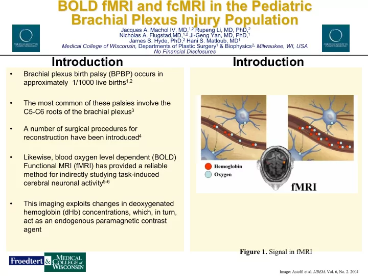
1. Foad SL, Mehlman CT, Ying J. The epidemiology of neonatal brachial plexus palsy in the united states. J Bone Joint Surg Am. 2008 Jun;90(6):1258-64. 2. Hoeksma AF, ter Steeg AM, Nelissen RG, van Ouwerkerk WJ, Lankhorst GJ, de Jong BA. Neurological recovery in obstetric brachial plexus injuries: An historical cohort study. Dev Med Child Neurol. 2004 Feb;46(2):76-83. 3. Narakas AO. Injuries of the brachial plexus and neighboring peripheral nerves in vertebral fractures and other trauma of the cervical spine. Orthopade. 1987 Feb;16(1):81-6. 4. Wood MB, Murray PM. Heterotopic nerve transfers: Recent trends with expanding indication. J Hand Surg Am. 2007 Mar;32(3):397-408. 5. Bandettini PA, Wong EC, Hinks RS, Tikofsky RS, Hyde JS. Time course EPI of human brain function during task activation. Magn Reson Med. 1992 Jun;25(2):390-7. 6. Kwong KK, Belliveau JW, Chesler DA, Goldberg IE, Weisskoff RM, Poncelet BP, et al. Dynamic magnetic resonance imaging of human brain activity during primary sensory stimulation. Proc Natl Acad Sci U S A. 1992 Jun 15;89(12):5675-9. 7. Fox M. D., Raichle M. E. (2007). Spontaneous fluctuations in brain activity observed with functional magnetic resonance imaging. Nat. Rev.

  • Neurosci. 8,

700–711. doi: 10.1038/nrn2201. 8. Biswal B, Yetkin FZ, Haughton VM, Hyde JS (1995) Functional connectivity in the motor cortex of resting human brain using echo-planar MRI. Magn Reson Med 34: 537–541. 8524021 9. Cordes D, Haughton VM, Arfanakis K, Carew JD, Turski PA, et al. (2001) Frequencies contributing to functional connectivity in the cerebral cortex in “resting-state” data. AJNR Am J Neuroradiol 22: 1326–1333.11498421 10. Li R, Machol JA,4th, Liu X et al. C7 nerve root sensory distribution in peripheral nerves: A BOLD fMRI investigation at 9.4T . Muscle Nerve. 2013. [Epub ahead of print] 11. Flugstad N, Stephenson J, Li R, Yan J, Hyde J, Matloub H. Cortical plasticity in a rat survival model of brachial plexus avulsion and cross C7 nerve transfer utilizing bold fMRI at 9.4 tesla. Plastic and Reconstructive Surgery. July 2012;130(1S):16. 12. Jones SR, Li R, Pawela CP, Shefchik DL, Matloub HS, Yan J-G, Jaradeh SS, Hyde JS. Cortical plasticity of the brain after median nerve transection using fMRI at 9.4T by direct nerve stimulation. Proc. Intl. Soc. Mag. Reson. Med. 2008 16: 2441. 13. Li R, Jones SR, Pawela CP, Shefchik DL, Yan J-G, Jaradeh SS, Matloub HS, Hyde JS. Functional MRI detection of acute and chronic brain plasticity following median and ulnar nerve transection using direct nerve stimulation at 9.4T Proc. Intl. Soc. Mag.

  • Reson. Med. 2008.

14. Parkins MA, Li R, Pawela CP, Matloub HS, Yan JG, Hyde JS. A peripheral nerve repair model using fMRI in rats. 17th Annual International Society for Magnetic Resonance in Medicine Meeting, Honolulu, HI. USA. 2009. 15. Pawela CP, Biswal BB, Hudetz AG, Li R, Jones SR, Cho YR, et al. Interhemispheric neuroplasticity following limb deafferentation detected by resting- state functional connectivity magnetic resonance imaging (fcMRI) and functional magnetic resonance imaging (fMRI). Neuroimage. 2010 Feb 1;49(3):2467

  • 78.

16. Huang RS, Sereno MI. Dodecapus: An MR-compatible system for somatosensory stimulation. Neuroimage. 2007 Feb 1;34(3):1060-73. 17. Anderson, A.W., Marois, R., Colson, E.R., Peterson, B.S., Duncan, C.C.,Ehrenkranz, R.A., Schneider, K.C., Gore, J.C., Ment, L.R., 2001.Neonatal auditory activation detected by functional magnetic resonance imaging.

  • Magn. Reson. Imaging 19, 1–5.

18. Nolte J. The Human Brain : An Introduction to its Functional Anatomy. Chapter 22 - Cerebral Cortex. Philadelphia, PA: Mosby Elsevier; 2009. 19. Cox RW, Hyde JS. Software tools for analysis and visualization of fMRI data. NMR Biomed. 1997 Jun-Aug;10(4-5):171-8.