
SLIDE 1
The Posterior Malleolus: Almost Always Fix it
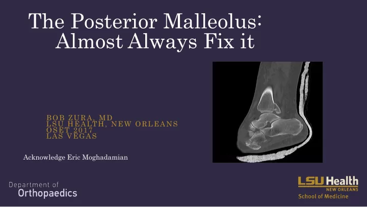
BOB ZURA, MD LSU HEALTH, NEW ORLEANS OSET 2017 LAS VEGAS
Acknowledge Eric Moghadamian
The Posterior Malleolus: Almost Always Fix it BOB ZURA, MD LSU - - PowerPoint PPT Presentation
The Posterior Malleolus: Almost Always Fix it BOB ZURA, MD LSU HEALTH, NEW ORLEANS OSET 2017 LAS VEGAS Acknowledge Eric Moghadamian Disclosures Consultant: Smith-Nephew Bioventus Cardinal Health Assumption Anatomic
BOB ZURA, MD LSU HEALTH, NEW ORLEANS OSET 2017 LAS VEGAS
Acknowledge Eric Moghadamian
– Smith-Nephew – Bioventus – Cardinal Health
errib ible le at diagnosing and reducing syndesmotic injuries
miss them
– Mostly PER injuries
Weening and Bhandari, 2005 Parikenen et al., 2011 JBJS
– But also in SER with no widening on static films:
Syndesmotic Instability in Weber B Ankle Fractures: A Clinical Evaluation Stark, Erik MD; Tornetta, Paul III MD; Creevy, William R MD
Journal of Orthopaedic Trauma 2007
manner, we often get it wrong.
– Incidence of malreduction based on CT scan “standard”: >50% – Gardner et al. FAI 27: 788-92, 2006.
syndesmotic injuries requiring reduction and fixation.
procedure underwent clinical and radiographic examination, computed tomographic (CT) scanning of both ankles (injured and uninjured)
Musculoskeletal Assessment and Olerud/Molander questionnaires. The Functional Consequence of Syndesmotic Joint Malreduction at a Minimum 2-Year Follow-Up Sagi, H. Claude MD; Shah, Anjan R. MD; Sanders, Roy W. MD
Journal of Orthopaedic Trauma Issue: Volume 26(7), July 2012, p 439–443
were available for follow-up.
translational asymmetry) when compared with the contralateral uninjured syndesmotic joint. The Functional Consequence of Syndesmotic Joint Malreduction at a Minimum 2-Year Follow-Up Sagi, H. Claude MD; Shah, Anjan R. MD; Sanders, Roy W. MD
Journal of Orthopaedic Trauma Issue: Volume 26(7), July 2012, p 439–443
malreduced on postoperative CT scans, whereas 44% (A/B) of the closed syndesmotic reductions were malreduced on postoperative CT scan (P = 0.11).
worse functional outcome scores (P < 0.05) on both the Short Form Musculoskeletal The Functional Consequence of Syndesmotic Joint Malreduction at a Minimum 2-Year Follow-Up Sagi, H. Claude MD; Shah, Anjan R. MD; Sanders, Roy W. MD
Journal of Orthopaedic Trauma Issue: Volume 26(7), July 2012, p 439–443
http://m8.i.pbase.com/o4/53/688553/1/92037578.lA9AXs4m.neworleans_pano_std.jpg
syndesmotic injuries demonstrated significantly worse functional
Olerud/Molander questionnaires.
lower rate of malreduction when evaluated by postoperative CT scan. The Functional Consequence of Syndesmotic Joint Malreduction at a Minimum 2-Year Follow-Up
Sagi, H. Claude MD; Shah, Anjan R. MD; Sanders, Roy W. MD Journal of Orthopaedic Trauma Issue: Volume 26(7), July 2012, p 439–443
perform a direct, open visualization of the syndesmosis during the reduction maneuver, but obtain a postoperative CT scan with comparison to the contralateral extremity as well.
must be given to revising the osteosynthesis. The Functional Consequence of Syndesmotic Joint Malreduction at a Minimum 2-Year Follow-Up
Sagi, H. Claude MD; Shah, Anjan R. MD; Sanders, Roy W. MD Journal of Orthopaedic Trauma Issue: Volume 26(7), July 2012, p 439–443
anatomic?
Clin Orthop Relat Res. 2006 Jun;447:165-71.
contribute the most to stability of the ankle syndesmosis
Stage 4 ankle fractures involving the posterior malleolus underwent MRI
fractures had complete PITFL ruptures
with posterior malleolus fx
simulate rigid fixation of a Weber C fibula fracture.
– 70% of intact stiffness
– 40% after using a syndesmotic screw.
historic controls with closed reduction
– Direct visualization for syndesmotic stabilization of ankle fractures. Miller AN, Carroll EA, Parker RJ, Boraiah S, Helfet DL, Lorich DG.Foot Ankle Int. 2009 May;30(5):419-26 – Posterior Malleolar Stabilization of Syndesmotic Injuries is Equivalent to Screw Fixation.Miller AN, Carroll EA, Parker RJ, Helfet DL, Lorich DG.Clin Orthop Relat
patients having syndesmotic screw fixation alone
address posterior malleolus