
SLIDE 1
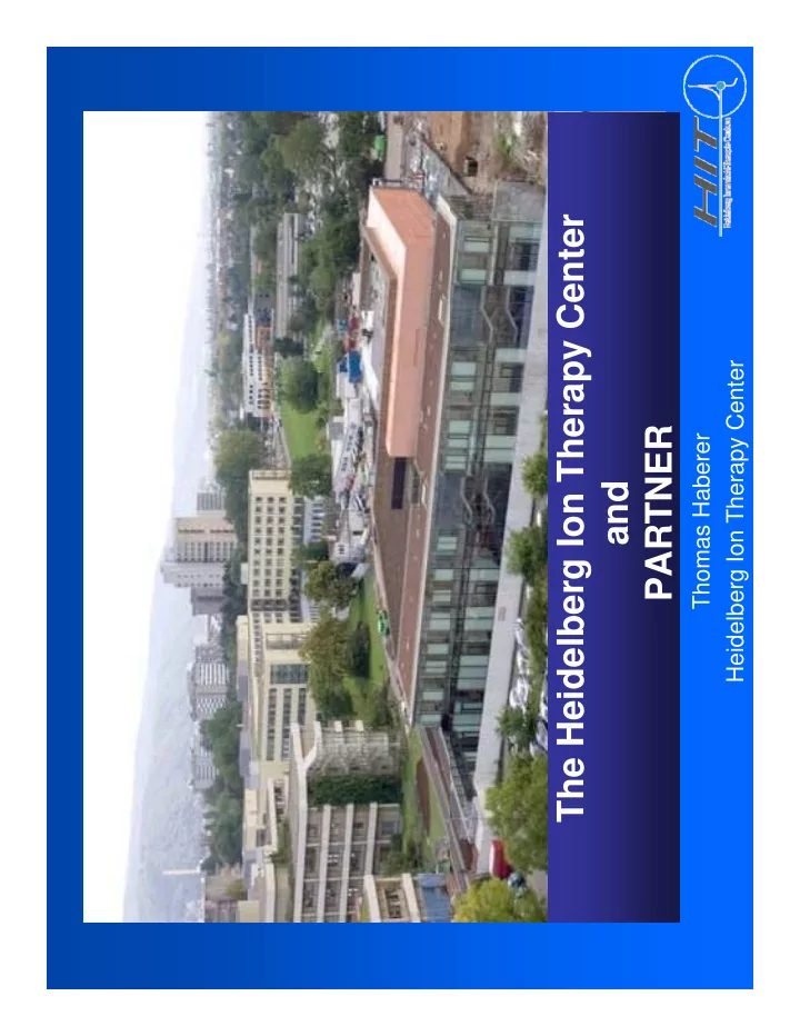
The Heidelberg Ion Therapy Center and PARTNER
Thomas Haberer Heidelberg Ion Therapy Center
The Heidelberg Ion Therapy Center Heidelberg Ion Therapy Center - - PDF document
The Heidelberg Ion Therapy Center Heidelberg Ion Therapy Center PARTNER PARTNER Thomas Haberer and Goal The key element to improve the clinical outcome is local control! local control! outcome is entrance channel: tumour: low
Thomas Haberer Heidelberg Ion Therapy Center
entrance channel:
tumour:
low rel. biol. effiency high rel. biol. effiency
g existing research accelerators Fi d hi
with moderate flexibility (if at all)
tumor-conform
(June 1994-August 2004)
Lung Pancreas Rectum 15 (1.2%) Eye 13 (1.0%) Miscell. 148 (11.4%) 245 (18.9%) Base of skull 20 (1.5%) 18 (1.4%)
Total 2,297
Head & Neck 207 (16.0%) Brain Esophagus 23 (1.8%) Uterus 78 (6.0%) Brain 74 (5.7%) Prostate 190 (14.6%) Liver 145 (11.2%) Bone/ soft tissue 121 (9.3%) ( )
scanning of focussed ion beams in fast in fast dipole magnets active variation
focus and intensity in the accelerator and accelerator and beam lines utmost precision via active position and intensity feed back loops back loops intensity-controlled rasterscan technique @ GSI Haberer et al., NIM A , 1993
253 energies (1mm range steps) x 7 spot sizes x 15 intensity steps
pp y
research
QA t
Pre OP
Post OP
dose [%]
Post OP t d b i
rasterscanned carbon ions
acute toxicity acceptable survival local control
Schulz-Ertner, Cancer 2005
y p late toxicity > CTC Grad 2 < 5%
full clinical integration
Protons (later He)
Carbon (Oxygen)
minutes ld id fi t
scanning ion gantry
> 15.000 fractions/year
7 500 tons Start of construction: November 2003 Completion of building and acc.: June 2006 First patient planned: early in 2009
p p y Project Partners: Project Partners:
GSI built the accelerator
related to patient environment GSI, DKFZ, … are research partners
: p
3He2+ 12C6 16O8+
: 48 72 88 102 (255 steps)
(255 steps)
: 4 - 10 mm (2d-gaussian) ( 4 steps)
RBE for fractionated RT of gut crypt cells of mice (Berkeley)
g yp ( y)
Proton data: Tepper et al. 1977, Ion data: Goldstein et al. 1981
RFQ + IH-DTL Ion sources
high energy beam transport synchrotron
Identical patient positioning systems
Workflow optimization
procedures
hand over from shuttle
I iti Inroom position verification
Open for future applications and workflows
preliminary scanner commissioning result Protons@maximum energy recorded in a verification film @ gy no feedback loops for beam intensity or position (courtesy S.O. Grözinger et al., Siemens Medical Solutions)
Advantage of a rotating beamline
Pancreas, supine position via gantry advantageous
application ld id fi t
ion gantry
integration
25m length 600to overall weight 0,5mm max. deformation
tested at GSI MT Mechatronics
MT Aerospace
tested at GSI MT Mechatronics
15 M1 Customisation and integration of the FLUKA MC code M6 Milestone: 15-M1 Customisation and integration of the FLUKA MC code in the UKL-HD (HIT) research planning platform for dose calculations of scanned ion beams in water and patient- or phantom-CTs M6 Milestone: Integration of the FLUKA Monte Carlo code in the research planning platform for planning platform for scanned ion beams at UKL-HD (HIT) 15-M2 15 D2 Experimental validation by means of dosimetric measurements in homogeneous and heterogeneous M12 Milestone: Experimental 15-D2 measurements in homogeneous and heterogeneous phantoms at UKL-HD (HIT) Experimental validation Deliverable: Report 15-D1 Development of workflow-efficient analysis tools for comparison of MC and analytical treatment plan calculations M18 Deliverable: Report 15 D2 Intercomparison between MC and analytical treatment M24 Deliverable: Report 15-D2 Intercomparison between MC and analytical treatment plan calculations in a representative number of challenging real clinical situations (e.g., in the presence
water/tissue ) M24 Deliverable: Report water/tissue…)
12C Reference Plan
Proton Plans ISO@140cm ISO@140cm ISO@80cm ISO@80cm
Calculations done with TRiP98 and TRiP98Beam (S B K P di) (S. Brons, K. Parodi)
2-M1 Effectiveness of carbon ions therapy m3 Specification 2-M2 Correct definition of treatment volumes & precise patient setup m6 Report 2-D1 Preliminary results: LC and early toxicity m6 Report 2-M3 Target definition for treatment m12 Protocol 2-M4 Technique for data analysis selected m21 Protocol 2-D2 Experimental data analysed m24 Report 2-D3 Preliminary results: LC, DFS, OS, and late toxicity and relationship with dose fractionation and Final Report m24 Report 2-D4 Clinical validation of biological input parameters m30 Report 2-D5 Cost-effectiveness analysis m36 Report
Courtesy S. Combs
2-M1 Refine epidemiological date for indication for ion therapy m6 Milestone 2-D1 Comparison of data on photon proton and ion therapy m12 Report 2 M2 D l li i l i l f ifi i di i 18 P l 2-M2 Develop clinical trial of specific indication m18 Protocol 2-D2 Report on the analysis of the data on P2-M2 m36 Report
Courtesy S. Combs
carbon ion therapy for chondrosarcomas at the base of the skull
local control 96.2% / 89.8% at 3 / 4 years, n=54 failure: n=1 in-field 5-year OS 98.2% failure: n=1 in-field n=1 border Courtesy S. Combs
Goals of this planned trial: Goals of this planned trial: Optimized therapy of chordomas and chondrosarcomas at the base of the skull at the base of the skull Comparison: high-dose proton vs carbon ion treatment, p g p , monitor localc control, overall survival and toxicity (phase III trial) Establish a common protocol Other trials are under way (pancreas, prostate, …)
Courtesy S. Combs, J. Debus, K. Herfarth, M. Münter
Courtesy by E. Rietzel (MGH) and C. Bert (GSI)
Organ motion + with beam scanning leads to interplay effects