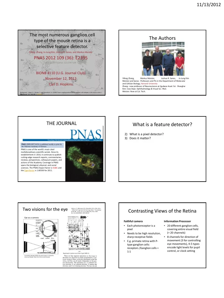

11/13/2012 The most numerous ganglion cell The Authors type of the mouse retina is a selective feature detector. Yifeng Zhang, In-Jung Kim, Joshua R. Sanes, and Markus Meister PNAS 2012 109 (36): E2395 doi/10.1073/pnas.1211547109 BIONB 4110 (U.G. Journal Club) November 12, 2012 Yifeng Zhang, Markus Meister, Joshua R. Sanes, In-Jung Kim Meister and Sanes: Professors and PIs in the Department of Molecular Carl D. Hopkins and Cellular Biology, Harvard University Zhang : now professor of Neuroscience at Cgubese Acad. Sci. Shanghai Kim: now Dept. Ophthalmology & Visual Sci. YALE. Meister: Now at Cal. Tech. Background: Volgyi, B., Chheda, S. and Bloomfield, S.A. (2009) Tracer coupling patterns of the ganglion cell subtypes in the mouse retina. J. Cop. Neurol. 512: 664-687. [PubMed] THE JOURNAL What is a feature detector? 2) What is a pixel detector? 3) Does it matter? PNAS is one of the world's most-cited multidisciplinary scientific serials. Since its establishment in 1914, it continues to publish cutting-edge research reports, commentaries, reviews, perspectives, colloquium papers, and actions of the Academy. Coverage in PNAS spans the biological, physical, and social sciences. The PNAS impact factor is 9.681 and the Eigenfactor is 1.60330 for 2011. Two visions for the eye Lettvin, J.Y.; Maturana, H.R.; Mcculloch, W.S.; Pitts, W.H.; Contrasting Views of the Retina , "What the Frog's Eye Tells the Frog's Brain," Proceedings of the IRE , vol.47, no.11, pp.1940-1951, Nov. 1959 doi: 10.1109/JRPROC.1959.287207 Eye as a camera Faithful camera Information Processor • Each photoreceptor is a • 20 different ganglion cells, pixel covering entire visual field • Needs to be high resolution, (= 20 channels) • 8 channels for direction of sharp receptive fields movement (3 for controlling • E.g. primate retina with P- eye movements), 4-5 types type ganglion cells: encode light levels for pupil receptors /Ganglion cells = control, or clock setting 1:1 Reprinted in Lettvin et al from Cajal 1909-11 from Wald, George (1953) ‘Eye and Camera’ in Scientific American Reader, New York, Simon and Schuster.
11/13/2012 The retina of has over 50 known cell types belonging to the 5 traditional classes (receptors, horizontal, bipolar, amacrine, ganglion) - mostly from Mouse Retina rabbit hotoreceptor orizontal ipolar macrine anglion From Masland (2001) Nature Neuroscience; reprinted in Gollisch and Meister Neuron, Volume 65, Issue 2, 150-164, 28 January 2010 Volgyi, B., Chheda, S. and Bloomfield, S.A. (2009) Tracer coupling patterns of the ganglion cell subtypes in the mouse retina. J. Cop. Neurol. 512: 664-687. [PubMed] 5 major cell types Principal Cells : the Ganglion cells (projection cells) axons go to • Supra-chiasmatic nucleus (circadian rhythms) • Pre-tectal nucleus (controls pupil size) • Superior colliculus (orients eye and head movements) • Lateral geniculate (form vision and movement) 5 major cell types
11/13/2012 Transgenic mouse lines permits selective How are the authors able to visualize only the W3 retinal labeling of RGC subtypes. Here, RGC W3 ganglion cell so that they can create a map of all of the W3 neurons in the retina? Transgenic mice Characteristics of W3 RGC • Th • Numerous • Small RF • High spatial resolution • Candidate pixel detector Thy-1 is a surface protein , a thymocyte antigen, used for marking stem cells, including axon processes of neurons. W3 expressing neurons in a mouse retina whole mount. Greater density in ventral retina. Left: vertical section of the inner plexiform layer. Red: ChAT label of amacrine cells (Choline acetyl transferase) Green: YFP label of W3 cells Blue: cell bodies (Nisl) Note: the dendrites of W3 are sandwiched between two bands of ChAT-positive bands in the IPL. Density of cells in the central retina cells/mm 2 Dashed- randomly spaced Red: hexagonal array
11/13/2012 The diameter of the dendritic arbor of W3 neurons is very small compared to other ganglion cells in the retina. The cell body is also small Spatial patterns can be: From Kim, Zhang, Meister, Sanes (2010) J. Neurosci. 30:1452 Coverage factor is 4.5 in densest region of retina (i.e 4 to 5 cells cover each point in space) W3 neurons are of a single cell type, with a relatively regular distribution. “Together , these results provide strong evidence that W3-RGCs are a single • The density recovery profile (density of W3 neurons versus their average spacing) cell type that represents the smallest-field and highest density RGCs in the indicates repulsion at close range; close fit to hexagonal packing. mouse retina. Hence one expects this population to sample the visual scene at the highest Expected if hexagonal spatial resolution .” Expected if random By using transgenic lines of mice containing YFP gene inserted into gene Thy1 get yellow fluorescence in subset of W3 RGC.
11/13/2012 Whole cell patch clamp from RGCs, some from W3 (guided by strong fluorescent label) others from non W3. Response to natural scene movies from “rat - cam”. Whole cell patch clamp from RGCs, some from W3 (guided by strong fluorescent label) Non-W3 RGCs respond to rat-cam movie; W3 cells are generally silent. others from non W3. Response to natural scene movies from “rat - cam”. Non-W3 RGCs respond to rat-cam movie; W3 cells are generally silent. Even though film shows a predatory bird flying nearby, the W3 neuron is silent, unless it’s receptive field is looking at the edge of the bird. How possible? – W3 does not encode Whole cell patch electrode records from one cell common visual events from moving or stationary objects. Natural stimuli • Recorded via head-mounted video (rat cam) • Contains significant pan motion due to angular and linear head movement – turning, walking • Presented to W3 cells – no response • For mouse in nature, head movements would be compensated by counter eye movement (VOR) but normal stimuli should contain significant optical flow patterns. Even though film shows a predatory bird flying nearby with a stationary background, the • What about when freezing? W3 neuron is silent, unless it’s receptive field is looking at the edge of the bird. How possible? – W3 does not encode common visual events from moving or stationary objects. The authors conclude that W3 neurons in the retina respond both to lights turning "on" and lights turning "off". How might this be possible, given that cones all respond to lights turning on with hyperpolarization?
11/13/2012 On-bipolar cells and off-bipolar cells terminate W3 Receptive Fields in different depths of the inner plexiform layer A. Shows the receptive field of the RGC overlaps with that of the dendritic arbor. Recording from W3, stimulate with\light, cell fires spikes on “ON” and on “OFF”. Averate spike rate for on vs. off responses plotted as a function of time. Note that the on response is delayed compared to the off response. WHY do you think this might be? B. Directionality of a moving bar stimulus. In each direction the cell fires on both edges of the bar (on, then off). Restricted receptive field area. C. Stimulus is a bnar of light and dark bands each fluctuating in intensity from light to dark. The spike-triggered stimulus average is determined, subjected to PCA, and represented as a two dimensional plot. The dots fall into two categories. Using PC1, the spike triggered reverse average is either excitatory followed yby inhibitory, or reversel The inhibition is faster and stronger. Spike triggered average, sorted by position in cluster shows that some spikes are driven by on, others by off. D. Model of the W3 ganglion cell is a single neuron with inputs from one On bipolar and one off bipolar, each is a rectifying synapse. The authors conclude that W3 retinal ganglion cells are "feature detectors" for arial predators. Make a list of characteristics of stimuli that cause the cells to fire that would lead to this conclusion. Voltage clamp data from W3 cell shows a series of responses at each holding potential. Global motion produced a large conductance increase, but note that the reversal potential is -61. Differential motion produced a weaker conductance incrase but the reversal potential is excitatory. Motion of the center grading alone produced a strong excitation, no inhibition.
11/13/2012 Response of W3 neuron to small dark objects such as a dark outline of a hawk flying Summary overhead on a bright background. Small objects, about the size of the W3 receptive field evoke the strongest response, independent of trace. No directional preference. A small, numerous, RGC, W3 is found over the entire retina. It has a small cell body, small, dense thorny dendritic field. W3 cells are a candidate for pixel detector in the retina. Transgenic lines selectively label W3 neurons with strong YFP signals. W3 neurons are silent to most stationary, and moving stimuli. W3 respond both to on and to off stimuli. Appear to receive both on and off bipolar inputs. Both are rectifying, and do not cancel each other out. In response to natural movie with head movements, cells do not respond. Other RGCs fire significantly. In response to moving stimuli appear to be strongly inhibited by peripheral movement. Inhibition is spike-dependent, from Amacrine cells. W3 neurons act as feature detectors for small dark objects on light background moving slowly.
Recommend
More recommend