
SLIDE 1
Structure-function analysis of the inositol trisphosphate receptor - - PDF document
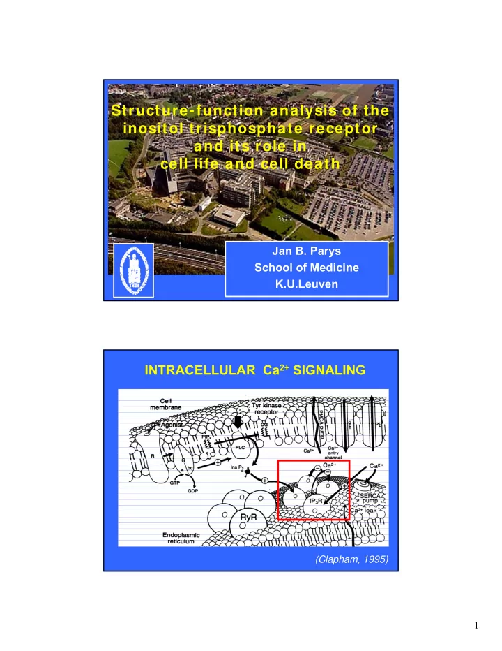
Structure-function analysis of the inositol trisphosphate receptor and its role in cell life and cell death Jan B. Parys School of Medicine K.U.Leuven INTRACELLULAR Ca 2+ SIGNALING (Clapham, 1995) 1 IP 3 R activation (Berridge, 2008) IP 3