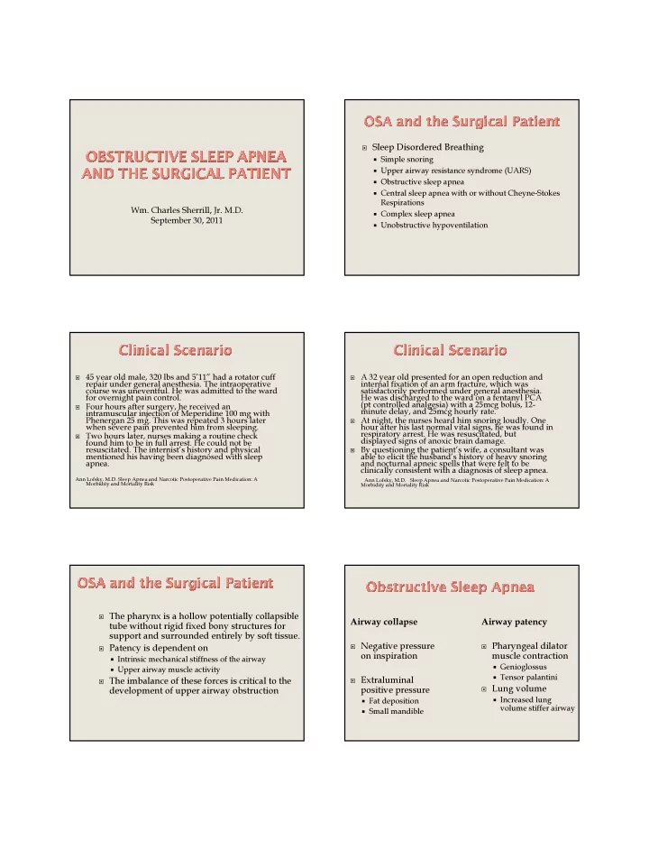

- Wm. Charles Sherrill, Jr. M.D.
September 30, 2011
Sleep Disordered Breathing Simple snoring Upper airway resistance syndrome (UARS) Obstructive sleep apnea Central sleep apnea with or without Cheyne-Stokes
Respirations
Complex sleep apnea Unobstructive hypoventilation
45 year old male, 320 lbs and 5’11” had a rotator cuff
repair under general anesthesia. The intraoperative course was uneventful. He was admitted to the ward for overnight pain control.
Four hours after surgery, he received an
intramuscular injection of Meperidine 100 mg with Phenergan 25 mg. This was repeated 3 hours later when severe pain prevented him from sleeping.
Two hours later, nurses making a routine check
found him to be in full arrest. He could not be
- resuscitated. The internist’s history and physical
mentioned his having been diagnosed with sleep apnea.
Ann Lofsky. M.D. Sleep Apnea and Narcotic Postoperative Pain Medication: A Morbidity and Mortality Risk A 32 year old presented for an open reduction and
internal fixation of an arm fracture, which was satisfactorily performed under general anesthesia. He was discharged to the ward on a fentanyl PCA (pt controlled analgesia) with a 25mcg bolus, 12- minute delay, and 25mcg hourly rate.
At night, the nurses heard him snoring loudly. One
hour after his last normal vital signs, he was found in respiratory arrest. He was resuscitated, but displayed signs of anoxic brain damage.
By questioning the patient’s wife, a consultant was
able to elicit the husband’s history of heavy snoring and nocturnal apneic spells that were felt to be clinically consistent with a diagnosis of sleep apnea.
Ann Lofsky, M.D. Sleep Apnea and Narcotic Postoperative Pain Medication: A Morbidity and Mortality Risk
The pharynx is a hollow potentially collapsible
tube without rigid fixed bony structures for support and surrounded entirely by soft tissue.
Patency is dependent on Intrinsic mechanical stiffness of the airway Upper airway muscle activity The imbalance of these forces is critical to the
development of upper airway obstruction Airway collapse
Negative pressure
- n inspiration
Extraluminal
positive pressure
Fat deposition Small mandible
Airway patency
Pharyngeal dilator
muscle contraction
Genioglossus Tensor palantini Lung volume Increased lung
volume stiffer airway