sarcoidosis pulmonary
play

Sarcoidosis: Pulmonary characteristics Manifestations, Diagnostic - PowerPoint PPT Presentation

Learning Objectives Have a better understanding of disease Sarcoidosis: Pulmonary characteristics Manifestations, Diagnostic How to make the diagnosis of sarcoidosis How to monitor patients Approaches and Treatment What is the


  1. Learning Objectives � Have a better understanding of disease Sarcoidosis: Pulmonary characteristics Manifestations, Diagnostic � How to make the diagnosis of sarcoidosis � How to monitor patients Approaches and Treatment � What is the natural history? Antonio D. Gomez, MD � What types of treatments are used? Assistant Professor of Medicine UCSF Online: Sarcoidosis.ucsf.edu Sarcoidosis: Disease Characteristics 
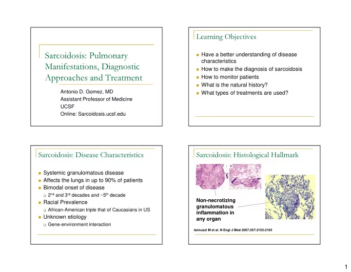
Sarcoidosis: Histological Hallmark � Systemic granulomatous disease � Affects the lungs in up to 90% of patients � Bimodal onset of disease � 2 nd and 3 rd decades and ~5 th decade Non-necrotizing � Racial Prevalence granulomatous � African-American triple that of Caucasians in US inflammation in � Unknown etiology any organ � Gene-environment interaction Iannuzzi M et al. N Engl J Med 2007;357:2153-2165 1

  2. Sarcoidosis: Clinical Presentations How to Make a Diagnosis of Sarcoidosis � Acute � Löfgren’s Syndrome � Fever, bilateral hilar lymphadenopathy, arthritis (ankle) No single diagnostic test! and erythema nodosum � Chronic � Subacute to chronic onset of symptoms � Often cough and/or shortness of breath � Systemic complaints in 25-50% � arthalgias, fatigue, chest pains, muscle pain ATS/ERS/WASOG. Am J Respir Crit Care Med. 1999 Costabel et al. Curr Opin Pulm Med. 2008 Diagnosis of Sarcoidosis Diagnosis of Sarcoidosis � Role of Angiotensin converting enzyme � High Resolution Chest CT scan (ACE) level � UCSF ILD radiologists think HRCT is very specific for the diagnosis if the classical patterns are present � Insensitive � Non-necrotic granulomas on tissue biopsy of � Non-specific affected organ � Elevated in other granulomatous diseases � Important to have an experienced lung pathologist review biopsy, especially if the interpretation is “granulomas with necrosis” ATS/ERS/WASOG. Am J Respir Crit Care Med. 1999 ATS/ERS/WASOG. Am J Respir Crit Care Med. 1999 2

  3. CXR Staging System Pulmonary Manifestations: Stage I, Bilateral Hilar Lymphadenopathy (BHL) Pulmonary Manifestations: Stage II, Pulmonary Manifestations: Stage III, BHL with Parenchymal Nodules Parenchymal Nodules Only Distribution: peri-lymphatic nodules, upper lobe nodules can coalesce � � 3

  4. Pulmonary Manifestations: Stage IV, Diagnosis of Sarcoidosis Fibrosis, Cystic � Exclusion of disease mimics: � Mycobacterial or fungal infection � Send tissue specimens for culture � Travel history (e.g. histoplasmosis, coccidioidosis) � Amyloidosis � Check SPEP and UPEP patients older than 50 or 60 in whom you are evaluating for sarcoidosis � Pneumoconiosis and Berylliosis � Take thorough occupational history � Lymphoma � Clinical history of B symptoms ATS/ERS/WASOG. Am J Respir Crit Care Med. 1999 Diagnosis of Sarcoidosis Diagnosis of Sarcoidosis � Sarcoidosis can be systemic � Tests that I don’t routinely perform � Gallium scans � Thorough review of systems Unless the patient cannot undergo tissue biopsy � � PET scans � May discover extrathoracic organ involvement Similar reasoning to gallium scans � E.g. skin, joints, cardiac, central or peripheral nervous � system � ACE levels Can consider if cannot obtain a biopsy � Sarcoidosis screening studies � � lysozme levels ATS/ERS/WASOG. Am J Respir Crit Care Med. 1999 4

  5. Sarcoidosis Screening Studies Monitoring Patients with Sarcoidosis � Pulmonary disease only: � Once diagnosis is made, the following screening is recommended: � Complete pulmonary function tests every 6 months during the first 2 years, and then yearly � 12 lead ECG and signal averaged ECG over the following 3 years unless symptoms � Serum Calcium level dictate for frequently � Precursor and mature forms of Vitamin D � Extrathoracic disease: � 25-hydroxy Vit D and 1, 25-dihydroxy Vit D � Depends on organ � Ophthalmologic evaluation � E.g Brain MRI for CNS sarcoidosis � 24 hr urine collection for calcium excretion � Monitor symptoms related to organ � Absolute CD4 count involvement ATS/ERS/WASOG. Am J Respir Crit Care Med. 1999 Natural History of Sarcoidosis Radiographic Staging: Predictor of Spontaneous Resolution � ~2/3 of patients have spontaneous resolution Spontaneous or persistent, but non-progressive disease Stage Chest Xray Finding Improvement I BHA 55-80% � ~1/3 have progressive disease II BHA, reticular infiltrates 40-60% � ~10% die from sarcoidosis-related organ involvement III Reticular infiltrates only 10-20% IV Fibrosis, volume loss 0% ATS/ERS/WASOG. Am J Respir Crit Care Med. 1999 5

  6. Treatment Recommendations Markers of Disease Activity First Assess the need for therapy � No clinically proven biomarkers of disease activity or progression Absolute indication for corticosteroids: � Cardiac* � ACE Level: should it be used to monitor � Neurologic* disease course? � Ophthalmologic � Not enough data to recommend routine use � Hypercalcemia � Some clinicians use ACE levels to assess disease activity � In patients who present with very elevated levels, it *Often high dose (60-80 mg/day) for first several may reflect disease activity weeks/months ATS/ERS/WASOG. Am J Respir Crit Care Med. 1999 Treatment Recommendations Treatment for Pulmonary Disease Progressive worsening of symptoms or PFTs � Non-life Threatening Disease or Severe Organ Dysfunction � ~ 40 mg prednisone for 3-6 wks, and if improved symptoms, taper by 5-10 mg � Expert opinion/Controversial topic increments every 4-8 wks � High rate of spontaneous remission and low mortality rate from pulmonary disease � Relapse rate can be up to 60%, so � Stage I pts (BHA) should be observed for 6 maintenance continued for 6-8 mos, resulting months and not treated in at least a year of treatment. � Early treatment of Stage II disease (BHA + � Second-line agents added for steroid- infiltrates) may improve lung function dependent, progressive disease or steroid � No data for disease-modification long term intolerance ATS/ERS/WASOG. Am J Respir Crit Care Med. 1999 ATS/ERS/WASOG. Am J Respir Crit Care Med. 1999 Pietinalho A. Chest. 2002 Pietinalho A. Chest. 2002 6

  7. Second-Line/Alternative Therapies Second-Line/Alternative Therapies � Methotrexate: (up to 15mg/week) � TNFa-blockers � DB-RCT: 15 new onset disease given MTX concordantly � Infliximab (Remicade): chimeric, humanized monoclonal with steroids. Less steroids used in MTX group. antibody Sarcoidosis Vasc Diffuse Lung Dis. 2000;17(1):60 � RCT: 138 patients with chronic pulmonary and extrapulmonary sarcoidosis refractory to glucocorticoid therapy (placebo, low- � Azathioprine (up to 200mg/day) dose (3 mg/kg), higher-dose (5 mg/kg) at baseline and weeks � open-label series studied azathioprine (2 mg/kg per day) 2, 6, 12, 18, and 24) combined with glucocorticoids in 11 patients with chronic or � Minimal improvement in FVC relapsing pulmonary sarcoidosis: Eur Respir J. � Adalimumab (Humira): fully human anti-TNFa antibody 1999;14(5):1117 � Case reports and small case series suggest benefit � Check serum thiopurine-S-methyltransferase (TPMT) to � Etanercept (Enbrel): soluble TNFa receptor fusion protein avoid severe pancytopenia � Not effective in pulmonary sarcoidosis Novel Therapies Take Home Points � IL-12 Antagonism (ustekinumab) � Sarcoidosis has a variable clinical course � Unpublished � Treatment often tailored to individual patient � All outcome measures negative � Spontaneous remissions are common � PRO � Difficult to predict progression, response to � Skin treatment, or relapse � Lung � No single test to indicate “active” disease � Treatment does not “cure” sarcoidosis 7

  8. Take Home Points: Treatment of GRADS: Genomics Research in Alpha-1 Pulmonary Disease Antitrypsin disease and Sarcoidosis � No evidence that any treatment is disease modifying � NIH funded, multicenter � ICS may be beneficial for cough without significant � UCSF -- only West Coast center radiographic disease � Study design: obtain clinical data, CT scans, � Methotrexate good for steroid-dependent and blood, and BAL specimens for genomic, refractory disease genetic and microbiomic analyses � Azathioprine good as second line after methotrexate � Goal: Identify markers for disease progression intolerance or treatment failure and determine the role of microorganisms in � Modest improvement with TNF- α inhibitors and disease etiology and progression very expensive � Enrollment: NOW Thank you for your attention! 8

Download Presentation
Download Policy: The content available on the website is offered to you 'AS IS' for your personal information and use only. It cannot be commercialized, licensed, or distributed on other websites without prior consent from the author. To download a presentation, simply click this link. If you encounter any difficulties during the download process, it's possible that the publisher has removed the file from their server.

Recommend


More recommend