
SLIDE 1
1
Pulmonary ABIM Certification Exam Review Course
Leslie Zimmerman, MD Professor of Clinical Medicine, UCSF ICU Director, SFVAMC
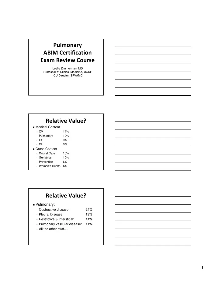
Relative Value?
Medical Content
- CV
14%
- Pulmonary
10%
- ID
9%
- GI
9%
Cross Content
- Critical Care
10%
- Geriatrics
10%
- Prevention
6%
- Women’s Health 6%
Relative Value?
Pulmonary:
- Obstructive disease:
24%
- Pleural Disease:
13%
- Restrictive & Interstitial:
11%
- Pulmonary vascular disease:
11%
- All the other stuff….