
SLIDE 1
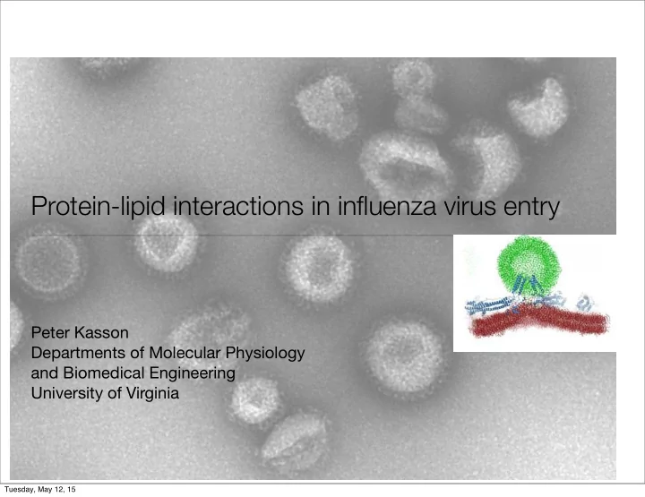
Protein-lipid interactions in influenza virus entry
Peter Kasson Departments of Molecular Physiology and Biomedical Engineering University of Virginia
Tuesday, May 12, 15
Protein-lipid interactions in influenza virus entry Peter Kasson - - PowerPoint PPT Presentation
Protein-lipid interactions in influenza virus entry Peter Kasson Departments of Molecular Physiology and Biomedical Engineering University of Virginia Tuesday, May 12, 15 Why is it hard to predict pandemics 1000+ 900+ 800+ 700+ Cases
Tuesday, May 12, 15
0+ 100+ 200+ 300+ 400+ 500+ 600+ 700+ 800+ 900+ 1000+ 1995+ 2000+ 2005+ 2010+ 2015+
Tuesday, May 12, 15
Viral membrane Target cell
Target and physical environment for fusion
FIGURE 8
Stages of fowl pl ague vi rus ent ry into MDCK cel ls . Ce l ls wi th prebound vi rus were warmed at 37°C for di f ferent t imes
and then f ixed wi th glutara ldehyde at room temperature . Wi thin 5 min, vi rus par t ic les were seen
in smooth sur faced pi ts and
vesic les (a, b, and c) , coated pi ts (d, e, and f) and coated vesic les (g, h, and
i )
. I nf, the samp l e was sta ined wi th ant i - fowl pl ague
vi rus spi ke prote in IgG and then wi th ferr i t in-goat ant i - rabbi t IgG af ter forma ldehyde f ixat ion (see Mater i a l s and Methods) . This
image demonst rates that par t of the vi rus par t icle was t ight ly assoc i ated wi th the membrane since onl y the exposed par t
is l abe l ed
wi th ferr i t in . Af ter 10 min, vi ruses were observed in endosomes ( j ) and mul t ivesi cul ar bodi es (k and I) . The images shown in
a, b,
and c were af ter 2 min warming ; in d, e, g,
k, and
i af ter 5 mi n warming, in f af ter 1 min warming, and in j , k, and / af ter 10 min
warming . a- i , x 62,500; k- l , x 50,000 .
brane of cel ls by lower ing the medium pH (5, 19, 54) . I f fowl plague vi rus infects MDCK cel ls by an endocytot ic pathway passing through the lysosomes,
i t might also be expected to
fuse at the plasma membrane i f exposed to low pH . This
seemed especial ly l ikely since low pH-dependent hemolysis and cel l -cel l fusion had been recent ly demonst rated for inf lu- enza vi ruses (19, 55, 56) . To test this, cel ls wi th prebound vi rus were suspended in media of pH 5 .0 and pH 7.4 for
1 min at
37°C and examined by t ransmission elect ron microscopy af ter
indi rect ferr i t in immunolabe l ing. In the cel ls kept at pH 7 .4, ferr i t in was associated only wi th vi rus part icles and not wi th the cel l sur face . No fusion of the vi rus wi th the cel l sur face was
In contrast ,
ferr i t in was at tached to the plasma
membrane only in samples exposed to low pH (Fig . 10) . In
several cases, clear cont inui ty between the cel l and vi rus mem-
branes was observed, wi th ferr i t in only bound to the prot ruding
vi rus prof i le (Fig . l0 a and b) . Fusion of vi ruses to membrane vesicles apparent ly shed f rom the cel ls was also observed (not
shown) . Low pH t reatment of MDCK cel ls in the absence of
vi rus produced some disturbance of the plasma membrane but did not induce art i factual ferr i t in binding .
To quant i tate low pH- induced fusion of fowl plague vi rus
to the MDCK cel l plasma membrane, cel ls wi th prebound radioact ive vi rus were incubated for 30 s at 37°C wi th media
FIGURE 10
Fus ion of fowl pl ague vi rus at the MDCK pl asma membrane . Fowl pl ague vi rus (60 j .g) was bound to MDCK cel ls for 1 h at 0°C and fusion was induced by incubat ing the cel ls for
1 min at pH 5 .0 and 37 °C. The cel ls were
then f ixed wi th forma ldehyde and immuno l abe l ed wi th ant i - fowl pl ague vi rus spi ke IgG and fer r i t in-conjugated goat ant i - rabbi t IgG . Vi ruses are
c lear ly recogni zabl e by the ferr i t in at tached to the i r membrane . In a the nuc l eocaps id
is st i l l c lear ly visible . In b the caps id is bare ly
recogni zabl e, but the vi rus shape and ferr i t in- labe led spi kes are st i l l obv ious
. In c the spi ke prote ins have presumabl y di f fused inthe pl ane of the membrane away f rom the si te of fusion . A vi rus prof i le
is st i l l detectabl e (ar row)
. Bar 0 .2 j m . x 106,000 .MATU i v
FT At .
Inf luenza Vi rus Ent ry
609
FIGURE 8
Stages of fowl pl ague vi rus ent ry into MDCK cel ls . Ce l ls wi th prebound vi rus were warmed at 37°C for di f ferent t imes
and then f ixed wi th glutara ldehyde at room temperature . Wi thin 5 min, vi rus par t ic les were seen
in smooth sur faced pi ts and
vesic les (a, b, and c) , coated pi ts (d, e, and f) and coated vesic les (g, h, and
i )
. I nf, the sampl e was sta ined wi th ant i - fowl pl ague
vi rus spi ke prote in IgG and then wi th ferr i t in-goat ant i - rabbi t IgG af ter forma ldehyde f ixat ion (see Mater i a ls and Methods) . This
image demonst rates that par t of the vi rus par t icle was t ight ly assoc i ated wi th the membrane since onl y the exposed par t
is l abe l ed
wi th ferr i t in . Af ter 10 min, vi ruses were observed in endosomes ( j ) and mul t ivesi cul ar bodi es (k and I) . The images shown in
a, b,
and c were af ter 2 min warming ; in d, e, g,
k, and
i af ter 5 min warming, in f af ter 1 min warming, and in j , k, and / af ter 10 min
warming . a- i , x 62,500; k- l , x 50,000 .
Matlin et al., 1981
Tuesday, May 12, 15
Waiting Time (s)
50 100 150 200
Num Fusion Events
5 10 15
Tuesday, May 12, 15
Tuesday, May 12, 15
Tuesday, May 12, 15
Tuesday, May 12, 15
Tuesday, May 12, 15
JACS 2011
Tuesday, May 12, 15
Pronk, Lindahl, Kasson. JACS 2015
5 10 15
Nw/Nl
5 10 15 20
<tp> / <tx>
with lipid diffusion correction without correction lipids single bilayer
Tuesday, May 12, 15
SIMD SIMD SIMD SIMD SIMD SIMD SIMD SIMD SIMD SIMD SIMD SIMD
Thread Thread Thread MPI MPI MPI Worker Worker Worker Server
IB SSL Shared memory
Average: 0.04MB/s Peak: 100MB/s Latency: 10 ms Average: 0.5GB/s Peak: >2.7GB/s Latency: 1-10μs Average: 0.5GB/s Peak: 25GB/s Latency: <100ns Cluster
Tuesday, May 12, 15
Tuesday, May 12, 15
Residue-Residue Contact Probability
181 186 191 196 201 206 211 216 181 186 191 196 201 206 211 216
0.1 0.2 0.3 0.4 0.5 0.6
time (us)
2 4 6 8 10 12
distance (nm)
0.5 1 1.5 2 2.5 3 3.5 4
Peptide-Peptide Center of Mass Distance : CG
Tuesday, May 12, 15
time (ns)
50 100 150
distance (nm)
0.5 1 1.5 2 2.5 3 3.5 4 4.5
Peptide-Peptide Center of Mass Distance : Quadruple Mutant
Tuesday, May 12, 15
Waiting Time (s) 50 100 150 200 Num Fusion Events 5 10 15
Tuesday, May 12, 15
Tuesday, May 12, 15