Pro: The Initial Treatment Should Always Be Debridement with Marrow - - PowerPoint PPT Presentation

pro the initial treatment should always be debridement
SMART_READER_LITE
LIVE PREVIEW

Pro: The Initial Treatment Should Always Be Debridement with Marrow - - PowerPoint PPT Presentation

Dalhosie University Halifax Nova Scotia Mark Glazebrook MSc., PhD, MD, FRCS(C), Dip Sports Med Associate Professor Dalhousie University Queen Elizabeth II health sciences Center Halifax, Nova Scotia Panel: 21-Year-Old College Junior Soccer


slide-1
SLIDE 1

MARK GLAZEBROOK MSc, PhD MD – Dalhousie Orthopaedics, CDHA, IWK, DGH

Mark Glazebrook

MSc., PhD, MD, FRCS(C), Dip Sports Med

Associate Professor Dalhousie University Queen Elizabeth II health sciences Center Halifax, Nova Scotia

Dalhosie University Halifax Nova Scotia

Pro: The Initial Treatment Should Always Be Debridement with Marrow Stimulation (6 minutes) Glazebrook MD PhD

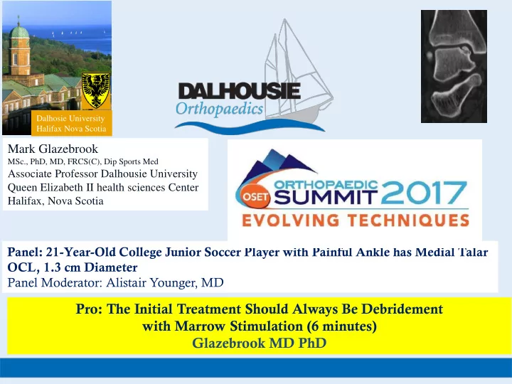
Panel: 21-Year-Old College Junior Soccer Player with Painful Ankle has Medial Talar OCL, 1.3 cm Diameter Panel Moderator: Alistair Younger, MD

slide-2
SLIDE 2

MARK GLAZEBROOK MSc, PhD MD – Dalhousie Orthopaedics, CDHA, IWK, DGH

Mark Glazebrook Disclosure Statement

Mark Glazebrook has received something of value in the past 1 year (≥ $500.00) or served as a Journal review er from a commercial company or institution related directly or indirectly to the subject of this presentation, as noted below.

a = research/institutional support, b = misc. non-income support, c = royalties, d = stock/options, e = consultant/employee f = Journal review er

NAME: DISCLOSURE: COMPANY/SOURCE: 1. Glazebrook e Stryker Wright Inc. 2. Glazebrook a,e Ferring Inc. 3. Glazebrook a,e Cartiva Inc 4. Glazebrook ae Smith & Nephew 5. Glazebrook f Foot & Ankle International 6. Glazebrook f JBJS(A) 7. Glazebrook f The Bone & Joint Journal 8. Glazebrook f CORR 9. Glazebrook Past BOD Member AOFAS

  • 10. Glazebrook

President Elect/BOD Canadian Orthopedics Association (COA)

slide-3
SLIDE 3

MARK GLAZEBROOK MSc, PhD MD – Dalhousie Orthopaedics, CDHA, IWK, DGH

Mark Glazebrook Disclosure Statement

Mark Glazebrook has received something of value in the past 1 year (≥ $500.00) or served as a Journal review er from a commercial company or institution related directly or indirectly to the subject of this presentation, as noted below.

a = research/institutional support, b = misc. non-income support, c = royalties, d = stock/options, e = consultant/employee f = Journal review er

NAME: DISCLOSURE: COMPANY/SOURCE: 1. Glazebrook e Stryker Wright Inc. 2. Glazebrook a,e Ferring Inc. 3. Glazebrook a,e Cartiva Inc 4. Glazebrook ae Smith & Nephew 5. Glazebrook f Foot & Ankle International 6. Glazebrook f JBJS(A) 7. Glazebrook f The Bone & Joint Journal 8. Glazebrook f CORR 9. Glazebrook Past BOD Member AOFAS

  • 10. Glazebrook

President Elect/BOD Canadian Orthopedics Association (COA)

slide-4
SLIDE 4

MARK GLAZEBROOK MSc, PhD MD – Dalhousie Orthopaedics, CDHA, IWK, DGH

Osteochondral Lesions

Definition and Terminology:

Osteochondritis Dissecans OCD (1887) Inflammatory process of bone cartilage Osteochodral Fracture (1959 Berndt & Harty) Primary Traumatic etiology Osteochondrosis Dissecans (1981) Repeated trauma or spontaneous or focal avascular osteonecrosis Octeochondral Lesions (OCL) Currently Used All encompassing

slide-5
SLIDE 5

MARK GLAZEBROOK MSc, PhD MD – Dalhousie Orthopaedics, CDHA, IWK, DGH

Osteochondral Lesions

Clinical: History

  • Persistent pain localized
  • Acute:
  • Pain swelling
  • Ecchymosis
  • Decreased range of motion
  • Chronic:
  • Dull ache
  • Stiffness
  • Crepitation
  • Tenderness
  • Mechanical clicking
  • Recurrent swelling
  • Locking or Instability……..
slide-6
SLIDE 6

MARK GLAZEBROOK MSc, PhD MD – Dalhousie Orthopaedics, CDHA, IWK, DGH

Osteochondral Lesions

Clinical: Physical

  • Swelling and ecchymosis
  • Localized Tenderness with palpation
  • Posterior dorsiflexion
  • Anterior plantarflexion
  • Range of motion:
  • stiffness
  • crepitus
  • clicking or locking.
slide-7
SLIDE 7

MARK GLAZEBROOK MSc, PhD MD – Dalhousie Orthopaedics, CDHA, IWK, DGH

Osteochondral Lesions

Clinical: Diagnostic Imaging

(Verhagen et al. 2005) Plain radiographs sensitivity - 0.70 specificity - 0.94 CT sensitivity - 0.81 specificity - 0.99 MR sensitivity - 0.96 specificity - 0.99 Arthroscopy sensitivity - 1.0 specificity - 0.97 CT SPEC Scan??????

slide-8
SLIDE 8

MARK GLAZEBROOK MSc, PhD MD – Dalhousie Orthopaedics, CDHA, IWK, DGH

Osteochondral Lesions

Clinical: Diagnostic Imaging

(Verhagen et al. 2005) Plain radiographs sensitivity - 0.70 specificity - 0.94 CT sensitivity - 0.81 specificity - 0.99 MR sensitivity - 0.96 specificity - 0.99 Arthroscopy sensitivity - 1.0 specificity - 0.97 CT SPEC Scan??????

slide-9
SLIDE 9

MARK GLAZEBROOK MSc, PhD MD – Dalhousie Orthopaedics, CDHA, IWK, DGH

Osteochondral Lesions

Classifications:

  • 1. Radiographic &/or Intra-op staging

Berndt and Harty 1959

  • 2. CT scan classification

Ferkel and Scranton 1993

  • 3. MRI classification

Hepple et al 1999

  • 4. Arthroscopic grading system

Pritsch 1986

slide-10
SLIDE 10

MARK GLAZEBROOK MSc, PhD MD – Dalhousie Orthopaedics, CDHA, IWK, DGH

OCD Talus: Classification

(Most recognized)

TABLE 1: BERNDT AND HARTY CLASSIFICATION

STAGE DESCRIPTION I Small subchondral compression II Incomplete fragment avulsion III Complete fragment detachment undisplaced IV Complete fragment detachment displaced

slide-11
SLIDE 11

MARK GLAZEBROOK MSc, PhD MD – Dalhousie Orthopaedics, CDHA, IWK, DGH

Osteochondral Lesions - Etiology Traumatic Etiology:

  • Lateral OCLs

Inversion, dorsiflexion & internal rotation  impaction fibula.

  • Medial OCLs

Inversion and plantar flexion  impaction of the posteromedial talar dome on tibia

Incidence of OCL in acute ankle injury

~ 70%

Coltart WD 1952, Bosien WR et al. 1955, Hintermann B et al. 200 and Leontaritis et al. 2009.

slide-12
SLIDE 12

MARK GLAZEBROOK MSc, PhD MD – Dalhousie Orthopaedics, CDHA, IWK, DGH

Osteochondral Lesions - Etiology Cystic Etiology (Van Dijk 2010) :

Atraumatic or traumatic etiology OCD lesions are increasingly associated with subchondral cysts.

  • Progression of simple non painful OCD’s to cystic

painful OCD’s is dependant on microfractured subchondral bone.

  • These subchondral defects allows fluid to be

compressed from cartilage into the subchondral bone leading to a localized high pressure

  • resultant local osteolysis causing a painful subchondral

cyst. The pain in osteochondral defects: Intermittent local rise in intraosseous fluid pressure with occurs on every step, and sensitizes the highly innervated subchondral bone.

From Van DIjk 2010

slide-13
SLIDE 13

MARK GLAZEBROOK MSc, PhD MD – Dalhousie Orthopaedics, CDHA, IWK, DGH

Osteochondral Lesions: Location

  • Anterolateral Common
  • Posteromedial Common
  • Central Uncommon
  • Posterolateral Uncommon

Difficult to access with standard anterior ankle arthroscopy

slide-14
SLIDE 14

MARK GLAZEBROOK MSc, PhD MD – Dalhousie Orthopaedics, CDHA, IWK, DGH

Surgical approach for Posteromedial OCL??

Anterior Posterior Combined Anterior & Posterior Approach Malleolar Osteotomy Trans tibial or Trans talar approach

slide-15
SLIDE 15

MARK GLAZEBROOK MSc, PhD MD – Dalhousie Orthopaedics, CDHA, IWK, DGH

12:00 1:00

Surgical approach for OCL

slide-16
SLIDE 16

MARK GLAZEBROOK MSc, PhD MD – Dalhousie Orthopaedics, CDHA, IWK, DGH

Level 1 Evidence………………..

Anterior Arthroscopy

12:00 1:00

Surgical approach for OCL

slide-17
SLIDE 17

MARK GLAZEBROOK MSc, PhD MD – Dalhousie Orthopaedics, CDHA, IWK, DGH

Level 1 Evidence………………..

Posterior Arthroscopy

12:00 1:00

Surgical approach for OCL

slide-18
SLIDE 18

MARK GLAZEBROOK MSc, PhD MD – Dalhousie Orthopaedics, CDHA, IWK, DGH

Combined Approach (Knee Flexed)

  • r

Flip Patient

12:00 1:00

Combined Approach (Knee Flexed)

  • r

Flip Patient

Surgical approach for OCL

Recommend

  • Dr. Ferkel’s Technique
slide-19
SLIDE 19

MARK GLAZEBROOK MSc, PhD MD – Dalhousie Orthopaedics, CDHA, IWK, DGH

Level 1 Evidence………………..

Anterior Arthroscopy Posterior Arthroscopy

Combined Approach (Knee Flexed)

  • r

Flip Patient

12:00 1:00

Combined Approach (Knee Flexed)

  • r

Flip Patient

Surgical approach for OCL

slide-20
SLIDE 20

MARK GLAZEBROOK MSc, PhD MD – Dalhousie Orthopaedics, CDHA, IWK, DGH

TREATMENT OF OCL

slide-21
SLIDE 21

MARK GLAZEBROOK MSc, PhD MD – Dalhousie Orthopaedics, CDHA, IWK, DGH

Treatment of OCL

Most published sources recommend non-operative treatment Stage I and II lesions It is generally agreed that operative intervention is recommended in Stage IV lesions and Stage I & II lesions with ongoing clinical symptoms Controversy exists for large stage III lesions and lesions in the skeletally immature patient.

slide-22
SLIDE 22

MARK GLAZEBROOK MSc, PhD MD – Dalhousie Orthopaedics, CDHA, IWK, DGH

Characteristics of an OCL require immediate OR Rx?

  • Limited Level 1 Evidence on Immediate Rx:
  • Type or Size

 Very Large & Cystic

  • Location

 Not a time sensitive Issue

  • Presence of Fragment  Flipped Fragment or Loose Catching
slide-23
SLIDE 23

MARK GLAZEBROOK MSc, PhD MD – Dalhousie Orthopaedics, CDHA, IWK, DGH

Clinical Outcomes of OCL Treatment

slide-24
SLIDE 24

MARK GLAZEBROOK MSc, PhD MD – Dalhousie Orthopaedics, CDHA, IWK, DGH

Evidence-Based Indications for Ankle Arth

Purpose : Review the literature available for the current generally accepted indications for ankle arthroscopy. Describe Level of Evidence (LOE) available to support generally accepted indications for ankle arthroscopy. Provide a Grade of Recommendation for Treatment based on LOE available.

Mark A. Glazebrook, Venkat Ganapathy, Michael A. Bridge, James W . Stone, Jean-Pascal Allard Arthroscopy: The Journal of Arthroscopic and Related Surgery December 2009 (Vol. 25, Issue 12, Pages 1478-1490)

slide-25
SLIDE 25

MARK GLAZEBROOK MSc, PhD MD – Dalhousie Orthopaedics, CDHA, IWK, DGH

Evidence Based Indication for Ankle Arthroscopy

Glazebrook, MA, Gonapathy V , Bridge M, Stone J, Amendola A, Allard JP Arthroscopy, Dec. 2009

Table 5 Summary of Grade of Recommendation for or against the current generally accepted indications for ankle arthroscopy. ___________________________________________________________ Procedure Grade of Recommendation ___________________________________________________________ Impingement (anterioer bone & soft tissue) B for

Osteochondral Defects (<15mm) B for

Ankle Arthrodesis B for Loose Bodies C for Ankle Instability C for Septic Arthritis C for Arthrofibrosis C for Ankle Osteoarthritis C against Fractures I for Synovitis I for

_______________________________________________

slide-26
SLIDE 26

MARK GLAZEBROOK MSc, PhD MD – Dalhousie Orthopaedics, CDHA, IWK, DGH

Summary of LOE for Arthroscopic Ankle OCL Rx

Level IV (20 case series)

  • no consensus on:

– grading of these lesions – best treatment modality

  • Outcomes ranged 33% to 100% good
  • Most studies (14) had over 80% good
  • utcome.
slide-27
SLIDE 27

MARK GLAZEBROOK MSc, PhD MD – Dalhousie Orthopaedics, CDHA, IWK, DGH

Summary of LOE for Arthroscopic Ankle OCL Rx

Level II-III (9)

  • Reports Trends:

– Good results 56-90% – Improved Clinical outcome scores (AOFAS) – Anterolateral better than Posteromedial – Cartilage removal better then penetration only – MRI and Arthroscopic grade not improved – No difference in primary vs previous failed surgery – Age not a limiting factor – Larger lesion (>1.5cm) may do better with advanced procedures

slide-28
SLIDE 28

MARK GLAZEBROOK MSc, PhD MD – Dalhousie Orthopaedics, CDHA, IWK, DGH

Ankle Arthroscopy well supported for Rx OCL.

(Grade B recommendation)

Arthroscopy and Osteochondral Lesions

slide-29
SLIDE 29

MARK GLAZEBROOK MSc, PhD MD – Dalhousie Orthopaedics, CDHA, IWK, DGH

Primary Treatment Non Tissue Transplantation Debridement with Bone Marrow Stimulation

slide-30
SLIDE 30

MARK GLAZEBROOK MSc, PhD MD – Dalhousie Orthopaedics, CDHA, IWK, DGH

Operative OCL Treatment Options

Non-tissue transplantation Primary internal fixation

  • -Excision/debridement
  • -Curettage
  • -Drilling
  • -Microfracture
slide-31
SLIDE 31

MARK GLAZEBROOK MSc, PhD MD – Dalhousie Orthopaedics, CDHA, IWK, DGH

OCD Debride Subchondral Penetration

  • Success Rates

75% -85% success Choi et al 2009 Am J Sports Med. 2009 Oct;37(10):1974-80 Zengerink et al Knee Surg Sports Traumatol Arthrosc. 2010;18( 2):238-46

75% of the time 75% Better….

Pre Operative 9 mos Post Operative

slide-32
SLIDE 32

MARK GLAZEBROOK MSc, PhD MD – Dalhousie Orthopaedics, CDHA, IWK, DGH

OCD Debride Subchondral Penetration

  • Success Rates

75% -85% success Choi et al 2009 Am J Sports Med. 2009 Oct;37(10):1974-80 Zengerink et al Knee Surg Sports Traumatol Arthrosc. 2010;18( 2):238-46

75% of the time 75% Better….

  • However up to 25% FAILURE

“RESISTENT OCLs” OR Size Matters???

Pre Operative 9 mos Post Operative

slide-33
SLIDE 33

MARK GLAZEBROOK MSc, PhD MD – Dalhousie Orthopaedics, CDHA, IWK, DGH

PURPOSE: Prognosis w ith defect size HYPOTHESIS: A critical, or threshold, defect size may exist at w hich clinical outcomes become poor in the treatment of osteochondral lesion of the talus Osteochondral lesion of the talus: is there a critical defect size for poor outcome? (Choi et al 2009) Am J Sports Med. 2009 Oct;37(10):1974-80

slide-34
SLIDE 34

MARK GLAZEBROOK MSc, PhD MD – Dalhousie Orthopaedics, CDHA, IWK, DGH

DESIGN:

  • Cohort study; Level III
  • 120 ankles underw ent arthroscopic marrow

stimulation treatment for OCL.

  • Clinical Failure Defined:

Reoperation AOFAS score less than 80 Osteochondral lesion of the talus: is there a critical defect size for poor outcome? (Choi et al 2009) Am J Sports Med. 2009 Oct;37(10):1974-80

slide-35
SLIDE 35

MARK GLAZEBROOK MSc, PhD MD – Dalhousie Orthopaedics, CDHA, IWK, DGH

RESULTS: 75% Success 25% (30 of 120 ) Clinical Failures:  18% (22) AOFAS <80  7% (8) required OATs Linear regression analysis show ed a high prognostic significance of defect area and suggested a cutoff defect size of 150 mm 2. CONCLUSION: Initial defect size is an important 150 mm 2. Osteochondral lesion of the talus: is there a critical defect size for poor outcome? (Choi et al 2009) Am J Sports Med. 2009 Oct;37(10):1974-80

slide-36
SLIDE 36

MARK GLAZEBROOK MSc, PhD MD – Dalhousie Orthopaedics, CDHA, IWK, DGH

Methods: (Level II) 105 patients prospective mean of 31.6 months and reported no treatment failures in lesions w ith a size Less than 1.5cm Microfracture for osteochondral lesions of the ankle: outcome analysis and outcome predictors

  • f 105 cases.

Chuckpaiwong et al. Arthroscopy. 2008;24( 1):106-12.

slide-37
SLIDE 37

MARK GLAZEBROOK MSc, PhD MD – Dalhousie Orthopaedics, CDHA, IWK, DGH

Treatment of Resistant Lesion? Advanced Procedure

  • r

Tissue Transplantation

slide-38
SLIDE 38

MARK GLAZEBROOK MSc, PhD MD – Dalhousie Orthopaedics, CDHA, IWK, DGH

Operative OCL Treatment Options

Non-tissue transplantation Primary internal fixation

  • -Excision/debridement
  • -Curettage
  • -Drillng
  • -Microfracture

Tissue transplantation “ADVANCED PROCEDURES” Osteochondral autograft transplantation

  • - Single plug
  • - Mosaicplasty

Osteochondral allograft transplantation Autologous chondrocyte transplantation Autologous stem cell transplantation Juvenile Cartilage Transplantation

slide-39
SLIDE 39

MARK GLAZEBROOK MSc, PhD MD – Dalhousie Orthopaedics, CDHA, IWK, DGH

Osteochondral lesions of the talus: randomized controlled trial comparing chondroplasty, microfracture, and

  • steochondral autograft transplantation

(Gobbi et al 2008) Arthroscopy. 2008 Feb;24(2):A16

  • METHODS:

– 33 Recalcitrant Ferkel class 2b, 3, and 4 OLT were randomized to:

  • 11 Chondroplasty
  • 10 Microfracture
  • 12 OATs

– Outcomes:

  • AOFAS
  • Subjective pain Assessment
  • MRI
slide-40
SLIDE 40

MARK GLAZEBROOK MSc, PhD MD – Dalhousie Orthopaedics, CDHA, IWK, DGH

Mean time to follow -up w as 53 months

  • Better outcome w as associated w ith smaller

lesions

  • All procedures similar clinical outcome scores

Injury Volume 39, Issue 1, Supplement

Osteochondral lesions of the talus: randomized controlled trial comparing chondroplasty, microfracture, and

  • steochondral autograft transplantation

(Gobbi et al 2008) Arthroscopy. 2008 Feb;24(2):A16

slide-41
SLIDE 41

MARK GLAZEBROOK MSc, PhD MD – Dalhousie Orthopaedics, CDHA, IWK, DGH

Autologous osteochondral grafting— Technique and long-term results (Hangody et al 2012) Arthroscopy. 2008 Feb;24(2):A16

  • METHODS: (Level IV)
  • 1097 pts OCD Rx w ith OATs
  • 98 talus
  • 93% good to excellent results by clinical
  • utcome scoring
  • 3% incidence of knee pain from donor site.
slide-42
SLIDE 42

MARK GLAZEBROOK MSc, PhD MD – Dalhousie Orthopaedics, CDHA, IWK, DGH

The Treatment of Osteochondral Lesions of the Talus with Autologous Osteochondral Transplantation and Bone Marrow Aspirate (Kennedy & Murawski 2009)

  • Results:
  • FAOS scores improved from 52.67 to 86.19
  • SF-12 scores also improved from 59.40 to 88.63
  • 3 patients (4 %) reported donor site knee pain

Conclusion: Autologous osteochondral transplantation is a reproducible and primary treatment strategy for large osteochondral lesion

  • f the talus.
slide-43
SLIDE 43

MARK GLAZEBROOK MSc, PhD MD – Dalhousie Orthopaedics, CDHA, IWK, DGH

My Approach

Size of Lesion only triggers me to counsel pts of outcome Scope Debride & Bone Marrow Stimulation for all to start Allows possible favorable outcome that has not been proven in quality studies to be more likely with advanced procedure (Gobbi et al 2008) Allows assessment of lesion to determine type of advanced procedure if necessary: Location Contained Size Bone quality

slide-44
SLIDE 44

MARK GLAZEBROOK MSc, PhD MD – Dalhousie Orthopaedics, CDHA, IWK, DGH

slide-45
SLIDE 45

MARK GLAZEBROOK MSc, PhD MD – Dalhousie Orthopaedics, CDHA, IWK, DGH

slide-46
SLIDE 46

MARK GLAZEBROOK MSc, PhD MD – Dalhousie Orthopaedics, CDHA, IWK, DGH

Arthroscopic Access Anterior?, Posterior ? or Combined??

12:00 1:00

slide-47
SLIDE 47

MARK GLAZEBROOK MSc, PhD MD – Dalhousie Orthopaedics, CDHA, IWK, DGH

slide-48
SLIDE 48

MARK GLAZEBROOK MSc, PhD MD – Dalhousie Orthopaedics, CDHA, IWK, DGH

Intact Cartilage????

slide-49
SLIDE 49

MARK GLAZEBROOK MSc, PhD MD – Dalhousie Orthopaedics, CDHA, IWK, DGH

Case 3 “Trampoline/Balotable” Cartilage

slide-50
SLIDE 50

MARK GLAZEBROOK MSc, PhD MD – Dalhousie Orthopaedics, CDHA, IWK, DGH

OCD with Mobile Fragment Excision

slide-51
SLIDE 51

MARK GLAZEBROOK MSc, PhD MD – Dalhousie Orthopaedics, CDHA, IWK, DGH

OCD Forage -Drilling

slide-52
SLIDE 52

MARK GLAZEBROOK MSc, PhD MD – Dalhousie Orthopaedics, CDHA, IWK, DGH

slide-53
SLIDE 53

MARK GLAZEBROOK MSc, PhD MD – Dalhousie Orthopaedics, CDHA, IWK, DGH

OCL Initial Rx Summary

  • Good History & Clinical Exam
  • Diagnostic Imaging CT Spec Preferred
  • Identify Large & Cystic Lesions to Counsel Pt.
  • Grade Lesion
  • I&II Non op first
  • III Non Op acute and Op Chronic
  • IV Operative
  • Surgery Location important for access
  • Scope Debride & Bone Marrow Stimulation for all to start
slide-54
SLIDE 54

MARK GLAZEBROOK MSc, PhD MD – Dalhousie Orthopaedics, CDHA, IWK, DGH

THANK YOU!!!