
SLIDE 1
1
Post-separation analysis
Pierre-Alain Binz Swiss Institute of Bioinformatics EMBNet course March 1-5, 2004
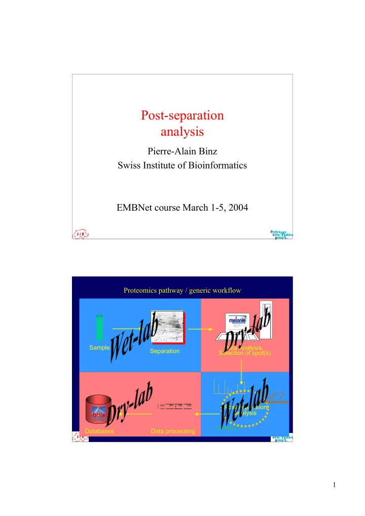
Proteomics pathway / generic workflow
Databases Separation Sample Data processing Data Analysis, Selection of spot(s)
G Q M R T N E K E
... NRTKGG ...
Post-separation analysis