
SLIDE 1
Phylogenetic analysis of Cytochrome P450 Phylogenetic analysis of - - PowerPoint PPT Presentation
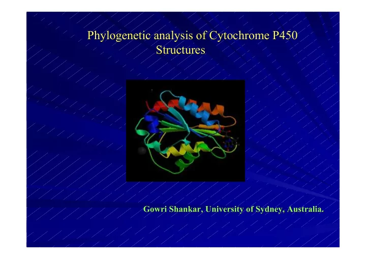
Phylogenetic analysis of Cytochrome P450 Phylogenetic analysis of Cytochrome P450 Structures Structures Gowri Shankar, University of Sydney, Australia. Gowri Shankar, University of Sydney, Australia. Overview Overview Introduction
Gowri Shankar, University of Sydney. Gowri Shankar, University of Sydney.
Gowri Shankar, University of Sydney. Gowri Shankar, University of Sydney.
Gowri Shankar, University of Sydney. Gowri Shankar, University of Sydney.
Gowri Shankar, University of Sydney. Gowri Shankar, University of Sydney.
Gowri Shankar, University of Sydney. Gowri Shankar, University of Sydney.
Gowri Shankar, University of Sydney. Gowri Shankar, University of Sydney.
Gowri Shankar, University of Sydney. Gowri Shankar, University of Sydney.
Gowri Shankar, University of Sydney. Gowri Shankar, University of Sydney.
Pseudomonas putida 1.6 20/07/2000 1dz4 Streptomyces coelictor 1.7 11/12/2202 1lfk Sulfolbus sulfataricus 1.5 28/02/2001 1io7 Mycobacterium tuberculosis 2.05 3/10/2003 1h5z Polyangium cellulosum 1.93 23/10/2003 1q5d Saccaropolyspora erythrea 2.1 28/04/2000 1eup Streptomyces coelictor 1.85 2/01/2004 1odo Fusarium oxysporum 1 20/12/2001 1jfb Pseudomonas Sp 2.3 26/11/1993 1cpt Thermus thermophilus 1.8 25/02/2003 1n97 Bacillus subtillis 2.1 18/03/2003 1izo Synthetic Pseudomonas putida 1.65 9/11/2001 1jpz Homo sapiens 2.05 27/07/2004 1tqn Oryctolagus cuniculus 1.6 7/10/2003 1po5 Oryctolagus cuniculus 2.1 12/08/2003 1nr6 Source Resolution Date PDB ID
Gowri Shankar, University of Sydney. Gowri Shankar, University of Sydney.
Gowri Shankar, University of Sydney. Gowri Shankar, University of Sydney.
Gowri Shankar, University of Sydney. Gowri Shankar, University of Sydney.
Gowri Shankar, University of Sydney. Gowri Shankar, University of Sydney.
SIMILAR Vs IDENTICAL AMINO ACID
5 10 15 20 25 30 35 40 45 50 15 25 35 45 55 65 75 SIMILAR IDENTICAL
Human with Human with Rabbit
Human with Archaea Human with Archaea
Gowri Shankar, University of Sydney. Gowri Shankar, University of Sydney.
500 1000 1500 1 2 3 4 5 6 7 DISTANCE DISTANCE
Human with Human with Rabbit
Human with Human with Archaea
Gowri Shankar, University of Sydney. Gowri Shankar, University of Sydney.
Gowri Shankar, University of Sydney. Gowri Shankar, University of Sydney.
Gowri Shankar, University of Sydney. Gowri Shankar, University of Sydney.
Gowri Shankar, University of Sydney. Gowri Shankar, University of Sydney.
Gowri Shankar, University of Sydney. Gowri Shankar, University of Sydney.
Gowri Shankar, University of Sydney. Gowri Shankar, University of Sydney.