
SLIDE 1
Sandro Olivo, Spokesperson, AXIm Group
(https://www.ucl.ac.uk/medphys/research/axim) Medical Physics and Bioemedical Engineering, UCL
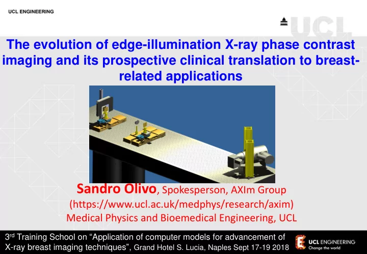
The evolution of edge-illumination X-ray phase contrast imaging and its prospective clinical translation to breast- related applications
3rd Training School on “Application of computer models for advancement of X-ray breast imaging techniques”, Grand Hotel S. Lucia, Naples Sept 17-19 2018