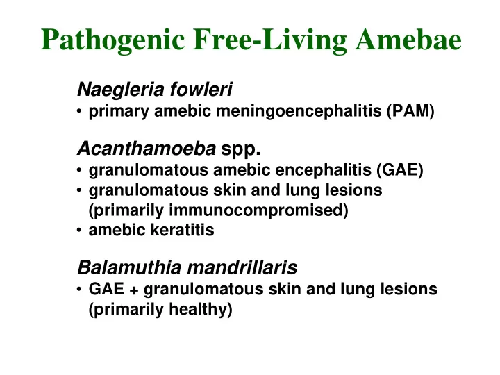
Pathogenic Free-Living Amebae Naegleria fowleri primary amebic - - PowerPoint PPT Presentation

pathogenic free living amebae
SMART_READER_LITE
LIVE PREVIEW

Pathogenic Free-Living Amebae Naegleria fowleri primary amebic - - PowerPoint PPT Presentation

Pathogenic Free-Living Amebae Naegleria fowleri primary amebic meningoencephalitis (PAM) Acanthamoeba spp. granulomatous amebic encephalitis (GAE) granulomatous skin and lung lesions (primarily immunocompromised) amebic keratitis


slide-1
SLIDE 1

Naegleria fowleri

  • primary amebic meningoencephalitis (PAM)

Acanthamoeba spp.

  • granulomatous amebic encephalitis (GAE)
  • granulomatous skin and lung lesions

(primarily immunocompromised)

  • amebic keratitis

Balamuthia mandrillaris

  • GAE + granulomatous skin and lung lesions

(primarily healthy)

Pathogenic Free-Living Amebae

slide-2
SLIDE 2

low nutrients desiccation

Naegleria Life Cycle

Cyst = dormant form feeding and replicating form Trophozoite =

slide-3
SLIDE 3

Naegleria fowleri

  • ubiquitous genus found in fresh water

lakes and ponds

  • PAM first recognized by Fowler (1965)
  • initially thought to be Acanthamoeba
  • Naegleria fowleri is only species

associated with PAM

  • ~ 200 documented cases worldwide
  • 81 in U.S.
  • 14 cases from same lake in Virginia
  • 16 cases from same stream feed pool in

Czech Republic

slide-4
SLIDE 4

Primary Amebic Meningoencephalitis (PAM)

  • 1-14 days incubation period
  • symptoms usually within a few days after

swimming in warm still waters

  • infection believed to be introduced through

nasal cavity and olfactory bulbs

  • symptoms include headache, lethargy,

disorientation, coma

  • rapid clinical course, death in 4-5 days after
  • nset of symptoms
  • trophozoites can be detected in spinal fluid, but

diagnosis is usually at autopsy

  • 4 known survivors treated with Amphotericin B
slide-5
SLIDE 5

brain section in vitro culture

“lobopodia”

slide-6
SLIDE 6

Acanthamoeba life cycle

slide-7
SLIDE 7
slide-8
SLIDE 8

Acanthamoeba

  • ubiqutous ameba of the soil and water
  • Culbertson (1958) fortuitously produced

disease in mice (culture contaminant)

  • human cases first reported in the early 70's
  • 73 cases worldwide of GAE as of 1991
  • 39 in U.S.
  • majority of patients are chronically ill,

immunocompromised, or debilitated with

  • ther diseases
  • also produces amebic keratitis and skin

and lung lesions

slide-9
SLIDE 9

Acanthamoeba Meningoencephalitis

  • portal of entry unknown, possibly respiratory tract,

eyes, skin

  • presumed hematogenous dissemination to the CNS
  • infection associated with debilitation or

immunosuppression

  • onset is insidious with headache, personality

changes, slight fever

  • progresses to coma and death in weeks to months
  • amebas not yet detected in spinal fluid
  • trophozoites and sometimes cysts detectable in

histological examination

  • no human cures documented
slide-10
SLIDE 10
slide-11
SLIDE 11

Amebic Keratitis

  • predisposing factors
  • ocular trauma
  • contact lens (contaminated cleaning solutions)
  • symptoms
  • ocular pain
  • corneal lesions (refractory to usual treatments)
  • diagnosis
  • demonstration of amebas in corneal scrapings
  • treatment
  • difficult, limited success
  • corneal grafts often required
slide-12
SLIDE 12

Balamuthia mandrillaris

  • first report in mandrill baboon (1990)
  • genus/species named 1993
  • morphology similar to Acanthamoeba
  • many Acanthamoeba GAE cases

retrospectively assigned to Balamuthia

  • as of 1997 63 cases of Balamuthia (30 in U.S.)
  • thus far only identified post-mortem
  • environmental source not yet identified
slide-13
SLIDE 13

Recavarren-Arce et al (Human Path. 30:269, 1999)

  • 10 autopsies (1985-97) of Balamuthia cases

in Peru

  • all healthy and all died within days or weeks
  • f neurological symptoms
  • primary lesions: 8 nasal, 3 dermal
  • questioned hematogenous dissemination in

both Acanthamoeba and Balamuthia

  • no intravascular ameba (this study and literature)
  • perivascular infiltration frequently observed
  • propose perivascular route from primary mucosal

lesion

slide-14
SLIDE 14

Red Tides

  • ‘blooms’ of dinoflagellates
  • phyto- and zooplankton
  • usually monospecific
  • rapid population increase
  • nutrients, lack of grazing,

sunlight, etc

  • long cyst survival in some

species

Increasing Incidence

  • better monitoring (seafood,

aquaculture)

  • coastal pollution/nutrients
  • agricultural wastes/fertilizers
  • long distance shipping
slide-15
SLIDE 15
  • deplete oxygen → dead zones
  • produce toxins → fish kills
  • accumulated dinoflagellate toxins pass

up the food chain

  • 'Ciguatera'
  • ‘shellfish’ poisoning

Potential Impact of Red Tides

Toxic Dinoflagellates and Shellfish Poisoning

Dinoflagellate Type of SP Toxin Gymnodinium breve neurotoxic brevetoxin Alexandrium tamerense paralytic saxitoxins Prorocentrum lima diarrhetic

  • kadaic acid

Pfiesteria piscicida neurotoxic + ?

slide-16
SLIDE 16

Pfiesteria piscicida

  • effects observed in late 1980’s (named 1996)
  • fisherman and swimmers complaining of rashes,

lesions, respiratory and neurological problems

  • massive fish kills in east coast estuaries
  • complex life cycle (at least 24 morphological forms)

Human Exposure to Pfiesteria Aerosols

  • narcosis/disorientation
  • respiratory distress/asthma-like
  • stomach cramping/nausea/vomiting
  • eye irritation/blurred vision
  • erratic heart beat (weeks)
  • sudden rages/personality changes
  • short term memory loss
slide-17
SLIDE 17

Why produce toxins?

  • culture filtrates induce open

ulcerative sores, hemorrhaging and death in fish

  • at least two toxins
  • heat-stable, water-soluble toxin

(fish become moribund within seconds and die within minutes)

  • lipophilic compound (causes the

epidermis to slough off)

Pfiesteria Toxins

  • accidental chemical affinity
  • self-defense (zooplankton, grazers)
  • ambush-predator life style
slide-18
SLIDE 18