
SLIDE 1
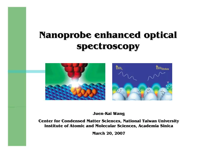
Nanoprobe enhanced optical Nanoprobe enhanced optical spectroscopy spectroscopy
J Juen uen-
- Kai Wang
Kai Wang Center for Condensed Matter Sciences, National Taiwan University Center for Condensed Matter Sciences, National Taiwan University Institute of Atomic and Molecular Sciences, Academia Sinica Institute of Atomic and Molecular Sciences, Academia Sinica March 20, 2007 March 20, 2007