
SLIDE 1
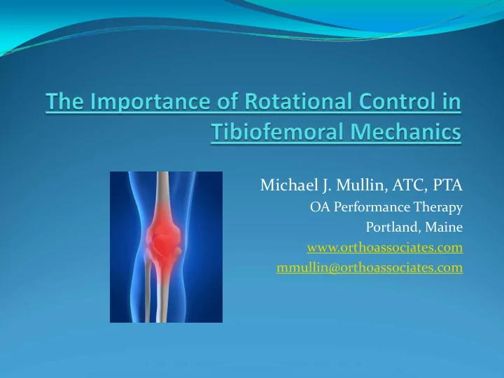
Michael J. Mullin, ATC, PTA
OA Performance Therapy Portland, Maine www.orthoassociates.com mmullin@orthoassociates.com
Michael J. Mullin, ATC, PTA OA Performance Therapy Portland, Maine - - PowerPoint PPT Presentation
Michael J. Mullin, ATC, PTA OA Performance Therapy Portland, Maine www.orthoassociates.com mmullin@orthoassociates.com Briefly review knee joint anatomy and arthrokinematics. Provide insight into new ways to evaluate the knee for
Michael J. Mullin, ATC, PTA
OA Performance Therapy Portland, Maine www.orthoassociates.com mmullin@orthoassociates.com
biomechanical contributing factors to pathology.
extremity control.
Osseous structures
Femur
MFC/LFC
Tibia
Lateral plateau‐‐convex Medial plateau‐‐concave
50% larger than lateral
Fibula Patella
facets
Meniscus
Lateral
Larger than medial More fully circular Consistent in width Greater mobility than
medial Medial
C‐shaped and broader
posteriorly than anteriorly
Attached to deep medial
capsule
Primary ligaments—varying
tension measures on different portions depending on joint position ACL PCL LCL—thin and round
Popliteus runs underneath
MCL—broad and flat
Superficial & deep fibers Deep fibers attach to medial
meniscus
Capsular and supporting
structures
Medial structures— viewed as 3 layers
retinaculum, fascial fibers of VMO, medial head gastroc
and gracilis near pes anserinus
Capsular and supporting
Lateral structures— divided into 3 layers
tendon
fabellafibular), capsule, popliteus
Other structures
Posterior capsule and
stabilizing ligaments and muscles.
Bursa Infrapatellar fat pad Plica Fascia
Muscular influences
mechanical forces producing strain, and/or structural / functional malalignment.
Knee extensors
Rectus femoris
Also flexes hip
Vastus intermedius Vastus lateralis Vastus medialis Patella tendon/ligament Articularis genus Iliotibial tract
In ranges between 0‐30
degrees
Knee flexors
Biceps femoris
(especially long head) Semimembranosus
Semitendinosis
Gastrocnemius
Iliotibial tract
Muscles influencing knee
Also function as stabilizers Popliteus
Medial/internal rotation (IR) Also unlocks knee from
terminal extension
Medial hamstrings
Medial rotation
Biceps femoris
Lateral/external rotation (ER)
movement and producing altered loads distally.
body past the point the pelvis and LE can stabilize effectively.
abduction)
Proper positioning of the hip and pelvis in a symmetrical pattern with no compensatory patterns further reduces strain onto
patterns of dysfunction in acetabulofemoral (AF) movement due to improper muscle sequencing.
Core control and the ability to isolate the deep TrA and pelvic floor muscles in varying positions dictates how the lower body will function during activity.
and distal joint position.
approximately 4 degrees results in considerably increased forces across the TF joint.
(“Musculoskeletal Biomechanics of the Knee
Orthopade. 2007 Jul;36(7):628‐34.)
During flexion, the femoral condyles roll posteriorly
During extension, the condyles roll anteriorly and
During knee flexion from an extended position, the
During knee extension, there is greater anterior
Every joint in the human body rotates or spins to some degree with movement (except the sutures of the skull). It is the ability to control excessive rotation and retraining the timing that is imperative in reducing joint and musculoskeletal strain. There are certain muscles which need to be inhibited while others that need to be facilitated in order to restore normal biomechanical control.
Muscular response
Popliteus, hamstrings, gracilis, sartorius, TFL/ITB, medial
head of gastroc
It is important to remember that the actions of most muscles
affecting movement change based on alterations in joint position proximally and/or distally. For example, the hamstrings increase their effectiveness as knee flexors as the hip moves into flexion and lose some as the hip moves into more extension. However, the motor control of the monoarticular muscles such as the popliteus and biceps femoris remain unchanged. Along these same lines is the role
effectiveness increases as the knee increases flexion closer to 90 degrees.
supination to pronation, and the tibia goes from stabilized ER to active IR, a lot of that motion is due to momentum. Some of it is active IR, but medial knee muscular and capsular tissue needs to stabilize; proximal and distal femur musculature needs to control excessive movement. Capsular structures take on a role of acting like “check reins” to reduce further movement.
alignment
anteversion
varus or valgus
ability of hip and pelvis stabilizers to allow for proper femoralacetabular and acetabular ‐femoral control.
femoral mobility in the joint will ultimately affect distal mechanics.
movement
restrictions
inflammation
considerations such as graft choice and mechancis of injury and subsequent surgery.
After Anterior Cruciate Ligament Reconstruction Using Ipsilateral Semitendinosus and Gracilis Tendon Autografts; Randall W. Viola, MD, William I. Sterett, MD, Darren Newfield, MD, J. Richard Steadman, MD and Michael R. Torry, PhD* ; Steadman Hawkins Clinic and Sports Medicine Foundation, Vail, Colorado. AJSM 28:552‐555 (2000).
movements
foot stance with one leg more horizontally abducted
patella”
posture
and/or increased lumbar lordosis
Ambulation
Increased apparent rotation throughout the lower
kinetic chain
Heel‐whip at toe‐off and swing phase of gait Poor ability to control pronation, especially barefoot Lateral shifting to one side more than contralateral or
“Sailor’s gait”
Functional movements Functional tests
Assessing and breaking down
certain movement patterns to further isolate source(s) of impairment aids in the development of a comprehen‐ sive treatment plan.
Looking for compensatory
movement (i.e. forward trunk position, side leaning to counterbalance, hip hiking, poor foot/ankle control)
Single leg balance Two‐legged squat or
Single leg squat
(bilaterally)
Bilateral jumping Unilateral hopping
Examination of TF rotational dysfunction
Observing other functional movements which may be contributing to their symptoms important as well—especially tasks which are repetitive.
and associated soft tissue restriction
patella
they pass the posteromedial tibia
insertions
misdiagnosed as ITBFS
Examination of TF rotational dysfunction Palpation‐Inf. Med. patella Palpation‐Inf. Med. patella
Examination of TF rotational dysfunction
Palpation—medial capsule and joint line
Examination of TF rotational dysfunction Palpation—pes anserinus and medial hamstrings
Examination of TF rotational dysfunction Palpation—popliteus at tibial and femoral insertions
Examination of TF rotational dysfunction Palpation—medial gastrocnemius head, popliteus, posterior medial structures
Seated OKC 90/90 ER/IR
Seated OKC 90/90 ER/IR
Supine circumduction palpation exam Beginning position Ending position
Supine circumduction palpation exam
One hand grasping the calcaneus while the other hand grasps joint line with
thumb on lateral and forefinger (or 2‐4 pads of fingers) along medial.
Starting with the hip and knee in fairly neutral positions. Begin with internally
rotating tibia while bringing hip into flexion and ER.
While cycling the knee through the motion, feel for the quality of movement of
the tibia on the femur. You should feel the medial tibial condyle drop posteriorly while the lateral condyle travels anterior and vice versa when bringing leg back into starting position.
Perform a number of times to feel where restrictions seem apparent, i.e. bands
Also feeling for restrictions in femoral rotation suggestive of altered loads
distally.
Soft tissue massage (STM) to the
medial → posterior capsule and joint line, beginning with more superficial tissue and working
direction of resistance.
STM to popliteus, medial
gastroc, and HS as needed. Popliteus is often thickened and therefore lost some contractile qualities.
Often referred to as strain/counterstrain Philosophy is to based on the theory that tissue can develop tone or a
shortened state in a specific location which can only be reduced by breaking the hyperactive (gamma gain) cycle. This is achieved by placing the affected tissue in a shortened state of comfort to reduce the tone (gamma efferent activity) and disrupting the dysfunctional position—essentially tricking it into submission.
While palpating the area of irritation, the practitioner moves bones to
manipulate the tissue into a shortened state which typically eliminates the point tenderness. This position is maintained and held for about 90 seconds. The body is then slowly taken out of this position which decreases the possibility that the affected tissue will return to its previous state.
Treatment of TF rotational dysfunction
techniques—MCL point/medial knee
leg off side of table and bent about 30 degrees, palpate the point of tone/pain at medial joint line, place a varus load
IR lower leg until tone is reduced. Hold 90 seconds, then slowly bring
Treatment of TF rotational dysfunction
Positional release techniques—ACL point/inferomedial fat pad Patient supine with towel roll under the distal femur, palpate point of tone/pain; lower leg held in IR by practitioner’s body then posterior glide of tibia with IR at the same time. 90 seconds, slowly release.
Treatment of TF rotational dysfunction
MWM’s for anterior talus—inability to squat fully or anterior ankle impingement with calf stretching, perform MWM’s to the talus focusing on posterior gliding as they actively PF and DF
Treatment of TF rotational dysfunction
Mobilization with movement techniques Moving tibia and femur through the range which was tested for dysfunction, the supine circumduction MWM.
Self mobilization techniques Grasp proximal tibia with leg in IR, shift weight forward and back while keeping IR pressure and femur going straight.
Self mobilization techniques
“Knee arounds”—kneeling with foot planted flat, pole placed in front of pinky toe, keeping foot flat, bring knee forward and around outside of pole, then back, making sure to keep weight from shifting laterally
Taping into IR—Patient standing with tibia in IR and femur in ER, start with tape at fibula head, grasp femur distally to pull into ER and pull tape medially. Pull around back of thigh and finish at lateral thigh.
Treatment of TF rotational dysfunction Taping into IR
Treatment of TF rotational dysfunction
Tibial IR factilitation—TB resisted tibial IR in seated 90/90 position
Glute med exercises as IR and abductor Sidelying with leg in IR
Lift into abduction and extension maintaining hip IR
External rotators as stabilizers
Clam exercise—starting position Clam exercise—finish position
Superclam Bridging with TB for ER
Treatment of TF rotational dysfunction
with TB wrapped around
while keeping foot flat and then rotate leg outwards against band. Come back to neutral, avoiding femoral IR. This shows set‐up position.
Half‐kneeling screen doors Starting position Half‐kneeling screen doors Finishing position
physioball with band around heel and another wrapped around both legs just below knees, flex knee while maintaining IR of tibia and
band at knees. Maintain good erect posture and avoid shifting weight laterally. This shows set‐up position.
TB resisted HS curls with IR Starting position TB resisted HS curls with IR Finishing position
Plank hip extensions Starting positions Plank hip extensions Finishing position
Plank hip extensions with knee flexion Starting position Plank hip extensions with knee flexion Finishing position
Squats with TB at knees and bolster at feet Starting position Squats with TB at knees and bolster at feet Finishing position
Lunges with TB resisted femoral IR Starting position Lunges with TB resisted femoral IR Finishing position
Tibial IR facilitation for poor horizontal abduction control with loading
Leg press with TB resistance Starting position (note foot position) Leg press with TB resistance Finishing position (note foot position)
Leg press with TB resistance Starting position (note changed foot position)
Leg press with TB resistance Finishing position (note changed foot position)
Supine HS bridge on physio with knee flex→ext (note ER foot position) Starting position
Supine HS bridge on physio with knee flex→ext (note ER foot position) Finishing position
Treatment of TF rotational dysfunction
careful instruction and monitoring of all exercises
benefit and avoidance of desired compensatory patterns.
position of feet with knees and heels separated but forefeet turned in towards each other. This IR is maintained throughout the exercise of extending hip and then going from flexion into extension and back.
TB resisted step‐ups Starting position TB resisted step‐ups Finish position
CKC glute wall exercise‐‐Starting position Maintain ER of femur into wall with squat CKC glute wall exercise‐‐Finish position Maintain ER of femur into wall with squat
Monster walks—maintain semi‐ squat position; do forward and back Monster walks—keep both legs from going into femoral IR when stepping
Trendelenburg jumps Starting position Trendelenburg jumps Finish position
rotational dysfunction.
soft tissue thickening, joint mobility restrictions, and try to manually normalize dysfunction.
pathology are identified and a treatment plan to address these is implemented. This includes educating the patient
the day. Make their day more symmetrical.
includes self mobilization techniques, careful instruction of an HEP and review of mechanics is essential.
Livingstone, New York, NY; 2000.
Frankel V. Lea & Febiger, Malvern, PA; 1989.