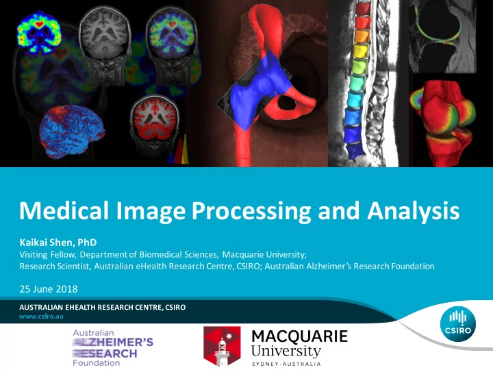

Medical Image Processing and Analysis Kaikai Shen, PhD Visiting Fellow, Department of Biomedical Sciences, Macquarie University; Research Scientist, Australian eHealth Research Centre, CSIRO; Australian Alzheimer’s Research Foundation 25 June 2018 AUSTRALIAN EHEALTH RESEARCH CENTRE, CSIRO
Medical Imaging and Image Analysis What we do • Develop and apply advanced computational tools to turn individual (and populations of) images into information (imaging biomarkers). – Accurate and reliable automated image analysis (reduces costs and may improve care), – provide new insights (Validity, reproducibility and predictive value), – enables new and improved diagnostics, screening and treatments. Medical Images Image analysis team Personalized medicine § Monitor individuals changes. § image processing § Patient specific treatments § registration § Characterize phenotype variability of disease. § segmentation § pattern recognition Preventing diseases § machine learning § Population studies § statistics § Early detection/screening § Diagnosis tools 2 | Medical Image Processing and Analysis | Kaikai Shen
Quantitative Image Analysis Overview of Neuroimaging Biomarkers we extract T1W T2W SWI DWI PET FLAIR Anatomy Venous tree White matter Amyloid beta load White matter CSF and structures connections Glucose metabolism lesions Neocortical Tissue atrophy Microbleeds White matter Connectivity uptake lesions strength Cortical thickness Iron deposit Patten of Axonal uptake Hippocampus volume integrity Atrophy patterns Tissue contrast 3 | Medical Image Processing and Analysis | Kaikai Shen
Quantitative Image Analysis Vertebrae Knee Prostate radiotherapy Hip planning with MRI 4 | Medical Image Processing and Analysis | Kaikai Shen
Structural MRI: structural changes in neurodegenerative diseases 5 | Medical Image Processing and Analysis | Kaikai Shen
Structural MRI: structural changes in neurodegenerative diseases Atrophy compared to Healthy control in early Alzheimer’s disease 6 | Medical Image Processing and Analysis | Kaikai Shen
Structural MRI: Volumetric Analysis Native Space Group-wise Skull Stripping Registration Segmentation Multi-Atlas (N=30) Multi-Atlas (N=15) Hippocampus Full brain Parcellation Parcellation 7 | Medical Image Processing and Analysis | Kaikai Shen
Structural MRI: Cortical thickness Meshing Cortical Thickness Smoothing and registration to template Z-score Web interface and report 8 | Medical Image Processing and Analysis | Kaikai Shen
Structural MRI (cont.) 9 | Medical Image Processing and Analysis | Kaikai Shen
CurAIBL: MR Assessment of Neurodegeneration https://milxcloud.csiro.au 10 | Medical Image Processing and Analysis | Kaikai Shen
FLAIR WM lesion segmentation 11 | Medical Image Processing and Analysis | Kaikai Shen
Diffusion MRI: Background Unrestricted diffusion Brownian motion 12 | Medical Image Processing and Analysis | Kaikai Shen
Diffusion MRI: Background Diffusion Tensor Imaging (DTI) 13 | Medical Image Processing and Analysis | Kaikai Shen
Diffusion MRI: Background Colour codes for orientation 14 | Medical Image Processing and Analysis | Kaikai Shen
Diffusion MRI: Background 15 | Medical Image Processing and Analysis | Kaikai Shen
Diffusion MRI: Background • Fibre Orientation Distribution (FOD) • Constrained spherical deconvolution (Tounier et al ., 2008) Tournier, J.-D., et al. , 2008. Resolving crossing fibres using Fibre Orientation Distribution constrained spherical deconvolution: Validation using diffusion- (FOD) weighted imaging phantom data. NeuroImage 42, 617–625. 16 | Medical Image Processing and Analysis | Kaikai Shen
Diffusion Tensor Imaging (DTI) 17 | Medical Image Processing and Analysis | Kaikai Shen
Fibre Orientation Distribution 18 | Medical Image Processing and Analysis | Kaikai Shen
single-shell single-tissue FOD ( b = 3000 s/mm 2 ) multi-shell multi-tissue FOD 19 | Medical Image Processing and Analysis | Kaikai Shen
FOD @ b = 3000 s/mm 2 multi-shell multi-tissue FOD 20 | Medical Image Processing and Analysis | Kaikai Shen
Genetic influence on connectivity • Aims • To develop new insights into brain development • To understand how our brains work in health, illness, youth, and old age • To study the cerebral cortex and the underlying neural connectivity, from the structural and diffusion MR images • To investigate the influence of genes by imaging monozygotic (MZ) and dizygotic(DZ) twins • Twin Study • Queensland Twin IMaging study (QTIM) • CSIRO and Queensland Institute of Medical Research (QIMR) de Zubicaray, G.I., Chiang, M.C., McMahon, K.L., Shattuck, D.W., Toga, A.W., Martin, N.G., Wright, M.J., Thompson, P.M., 2008. Meeting the challenges of neuroimaging genetics. Brain Imaging Behav. 2, 258–263. 21 | Medical Image Processing and Analysis | Kaikai Shen
Genetic influence on connectivity: Methods • Processing/Analysis framework • Measure the FODs • Raffelt et al ., 2012 • Peak amplitude FOD peak amplitude Group average FOD of average FOD template in the common space (b) (c) Iterateive Find peaks Groupwise registration (a) (d) Preprocessing Find peaks on FOD and spherical images corresponding deconvolution Fibre Orientation Diffusion Weighted to the peaks of the FOD peak amplitude Distribution (FOD) Images group average FOD of FOD images (e) Registered FOD images Inter-subject in the common space 1 st peak statistical analysis 2 nd peak Test-retest Heritability of reliability of FOD peak FOD peak measure measure Raffelt, D., et al ., 2012. Apparent Fibre Density: A novel measure for the analysis of diffusion-weighted magnetic resonance images. NeuroImage 59, 3976–3994. 22 | Medical Image Processing and Analysis | Kaikai Shen
Genetic influence on connectivity: Methods • Diffusiton MR: 94 gradient directions at b = 1159 s/mm 2 • Twin cohort • N =328 subjects (118M, 210F), age 22.7(2.3) • 71 pairs ( N =142, 48M, 94F) of monozygotic twins (MZ) + 90 pairs ( N =180, 69M, 111F) of dizygotic twins (DZ) • Heritability • ACE model: Additive genetics + Common environment + unique Environment FOD = A + C + E Falconer’s formula • Heritability h 2 = 2( r MZ – r DZ ) Var( A ) 2 h = Var( A ) Var( C ) Var( E ) + + 23 | Medical Image Processing and Analysis | Kaikai Shen
24 | Medical Image Processing and Analysis | Kaikai Shen
Genetic influence on connectivity: Methods • Tractography • Whole brain, probabilistic using FOD • Tract-wise heritability • Interpolation of heritabilities of nearest peaks • Tract average heritability 25 | Medical Image Processing and Analysis | Kaikai Shen
Human Brain Mapping Volume 37, Issue 6, pages 2331-2347, 23 MAR 2016 DOI: 10.1002/hbm.23177 http://onlinelibrary.wiley.com/doi/10.1002/hbm.23177/full#hbm23177-fig-0004 26 | Medical Image Processing and Analysis | Kaikai Shen
Heritability maps of cortical thickness. From left to right: heritability index h 2 , intraclass correlation between monozygotic twins ICC MZ , intraclass correlation between dizygotic twins ICC DZ . 27 | Medical Image Processing and Analysis | Kaikai Shen
The genetic correlation r g between the cortical thickness and white matter connectivity measured for each cortical region. 28 | Medical Image Processing and Analysis | Kaikai Shen
Positron Emission Tomography (PET) • β-amyloid plaque in Alzheimer’s disease • PET 11 C-PiB has been used as the tracer in many clinical studies since 2006 29 | Medical Image Processing and Analysis | Kaikai Shen
PET: Quantification Multi-atlas - Local Patch based selection. - Bayesian Fusion - Estimated GM Results: MR based top PET only bottom Zhou et al . ( PloS one ; Jan, 2014: DOI: 10.1371/journal.pone.0084777) 30 | Medical Image Processing and Analysis | Kaikai Shen
CapAIBL: PET Assessment of Neurodegeneration MILXCloud: https://milxcloud.csiro.au/ • CapAIBL: PET quantification • CurAIBL: MRI 31 | Medical Image Processing and Analysis | Kaikai Shen
Acknowledgement Olivier Salvado Prof. Ralph Martins Jurgen Fripp (MQ, AARF) (CSIRO) Sabine Bird Pratishtha Chatterjee Belinda Brown Pierrick Bourgeat Cintia Dias Samatha Gardener Shekhar Chandra Mitra Elmi Simon Laws Amy Chan Sunil Gupta Stephanie Rainey-Smith David Conlan Maryam Mohamaadi Hamid Sohrabi Vincent Doré Danit Saks Kevin Taddei Amir Fazlollahi Tejal Shah Sherilyn Tan Aleš Neubert Michael Weinborn Kerstin Pannek Anthony Paproki Parnesh Raniga Lee Reid Ying Xia 32 | Medical Image Processing and Analysis | Kaikai Shen
Thank you Kaikai Shen Kaikai.Shen@csiro.au AUSTRALIAN EHEALTH RESEARCH CENTRE 33 | Medical Image Processing and Analysis | Kaikai Shen
Recommend
More recommend