March 3 rd and 5 th , 2010 Biochemistry Recitation MBioS 303 - - PDF document

march 3 rd and 5 th 2010 biochemistry recitation
SMART_READER_LITE
LIVE PREVIEW

March 3 rd and 5 th , 2010 Biochemistry Recitation MBioS 303 - - PDF document

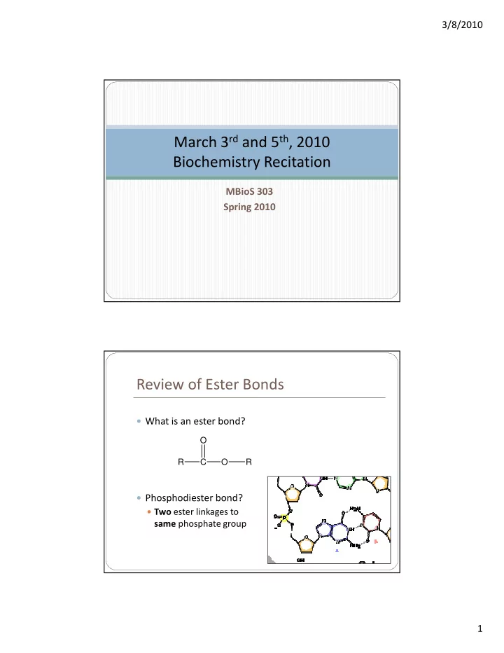
3/8/2010 March 3 rd and 5 th , 2010 Biochemistry Recitation MBioS 303 Spring 2010 Review of Ester Bonds What is an ester bond? O R C O R Phosphodiester bond? Two ester linkages to same phosphate group 1 3/8/2010 Review of


slide-1
SLIDE 1

3/8/2010 1 MBioS 303 Spring 2010

March 3rd and 5th, 2010 Biochemistry Recitation Review of Ester Bonds

What is an ester bond? Phosphodiester bond?

Two ester linkages to

same phosphate group

C O O R R

slide-2
SLIDE 2

3/8/2010 2

Review of Ester Bonds

What is a phosphoanhydride bond?

High-energy bond between two phosphate groups

γ β α γ β α γ β α γ β α

QUIZ 4

slide-3
SLIDE 3

3/8/2010 3

Outline

DNA Base Pairing and Tm Structural Features (palindromes, hairpins, etc.) DNA Damage DNA Supercoiling DNA Sequencing Higher Order DNA Structure (Chromatin)

DNA Base Pairing and Tm

Biochemistry, 5th Ed.

What DNA bases typically pair with each other?

slide-4
SLIDE 4

3/8/2010 4

DNA Base Pairing and Tm

Molecular Cell Biology, W.H. Freeman & Co., 1999

How would you define Tm?

DNA/RNA Structural Features

slide-5
SLIDE 5

3/8/2010 5

DNA Structural Forms

A-DNA: More compact than B; found in dessicated forms B-DNA: Structure typically found in cells. KNOW the above characteristics! Z-DNA: More stretched than B; the only left-handed form.

DNA Structural Forms

Which pictures represent A, B and Z DNA?

A B C A B C

Genomes, 2nd Ed.

slide-6
SLIDE 6

3/8/2010 6

DNA Structural Forms: Palindromes

Both strands have the same sequence, in antiparallel

  • rientation

Examples: GAATTC ACCTAGGT CTTAAG TGGATCCA GACTCCXXXXXXGGAGTC CTGAGGXXXXXXCCTCAG

http://en.wikipedia.org/wiki/Image:Stem-loop.svg Landes Bioscience, Eurekah.com

See example

  • n board

Mirror Repeats

The same sequence is present on one strand, as a

mirror image

Can’t form hairpins

slide-7
SLIDE 7

3/8/2010 7

Mirror Repeats

The same sequence is present on one strand, as a

mirror image

Can’t form hairpins CAN form DNA triple helices Functional importance: produces sharp bend in DNA

Landes Bioscience, Eurekah.com

DNA Damage

slide-8
SLIDE 8

3/8/2010 8

Who Cares?

Damaged RNA and proteins can be easily replaced by

reading from the DNA sequence

There is no way to replace the DNA molecules! Unrepaired DNA mutations linked to disease, including

cancer

Unrepaired mutations may be passed to offspring if

  • ccur in gametes

Cells have a number of DNA repair systems to fix such

mutations

Sources of Damage

Endogenous cellular sources

Reactive Oxygen Species (ex. from electron transport

chain)

Base hydrolysis (ex. deamination, depuriniation, etc.) Base mismatch (from errors in DNA replication)

Exogenous sources of damage

UV light Ionizing radiation Chemical sources (may cause many types of DNA

damage)

slide-9
SLIDE 9

3/8/2010 9

Depurination

Hydrolysis of N-glycosidic bond linking sugar and purine

base

Occurs spontaneously in the cell

Up to 10,000 purines lost from DNA every 24 hours in a mammalian

cell

The Cell: A Molecular Approach, 2nd Ed.

What kind of problem could this cause in a cell? Why?

Deamination

Loss of exocyclic amino

groups

Occurs spontaneously in

the cell

About 100 deaminations

  • ccur in a mammalian

cell every day

May explain why DNA

contains T rather than U

The Cell: A Molecular Approach, 2nd Ed.

slide-10
SLIDE 10

3/8/2010 10

UV Induced DNA Damage: CPDs

UV light

Cross-linking between C5 and C6 on adjacent pyrimidine

bases (usually thymine) forming a ring structure = cyclobutane pyrimidine dimer (CPD)

Molecular Cell Biology, W.H. Freeman & Co., 1999

UV Induced DNA Damage: CPDs

Causes “kink” in DNA Dimer can’t fit

normally into double helix, blocking replication and gene expression until removed

http://home.twcny.rr.com/geomanagement/ensmingr/menagerie.html

slide-11
SLIDE 11

3/8/2010 11

UV Induced DNA Damage: 6-4 PP

Cross-linking between C6 on one

pyrimidine to C4 on an adjacent pyrimidine = 6-4 photoproduct

Occur less frequently than CPDs,

but are more damaging

Cause bigger “kink” in DNA,

which tends to be lethal rather than mutagenic

Modern Genetic Analysis

DNA Damage: Summary

Deamination Depurination Cyclobutyl pyrimidine dimers 6-4 photo- products

Cause Spontaneous Spontaneous UV light UV light Result Loss of exocyclic amine group Loss of purine base Formation of cyclobutyl ring structure; can block replication and gene expression Formation of 6- 4 crosslink that causes significant kink in DNA backbone; can block replication/ gene expression

slide-12
SLIDE 12

3/8/2010 12

DNA Supercoiling Supercoiling

Coiling a coil Intrinsic property of

DNA

slide-13
SLIDE 13

3/8/2010 13

Linking Number

Lk = number of times

  • ne strand passes

through the “plane” created by the other strand

Change in Linking Number

For B-DNA, Linking

number (Lk) = (# base pairs)/10.5

More base pairs per

turn (ex. 12 instead of 10.5) = -Lk

Fewer base pairs per

turn (ex. 9 instead of 10.5) = +Lk

slide-14
SLIDE 14

3/8/2010 14

Effect of Fixed DNA Ends

Molecular Biology of the Cell, 4th Ed.

Function of Topoisomerases?

Molecular Biology of the Cell, 4th Ed.

slide-15
SLIDE 15

3/8/2010 15

Topoisomerase I

Releases strain from over- or under-winding of the DNA Forms a single-stranded break, and changes linking

number by 1

ATP independent

Molecular Cell Biology, 4th Ed.

Topoisomerase II (ex. DNA Gyrase)

Releases strain from underwinding of the DNA Forms a double-stranded break, and changes linking

number by 2

ATP dependent

Molecular Cell Biology, 4th Ed.

slide-16
SLIDE 16

3/8/2010 16

Comparison of Topoisomerases

Topoisomerase I Topoisomerase II Mechanism single-strand break double-strand break Change in Lk 1 2 Reliance on ATP ATP independent ATP dependent