late presentation of anomalous origin of the left
play

Late presentation of anomalous origin of the left coronary artery - PDF document

Case Report http://www.alliedacademies.org/annals-of-cardiovascular-and-thoracic-surgery/ Late presentation of anomalous origin of the left coronary artery from the pulmonary artery (ALCAPA) was confused with coronary artery fistula. Bashir A


  1. Case Report http://www.alliedacademies.org/annals-of-cardiovascular-and-thoracic-surgery/ Late presentation of anomalous origin of the left coronary artery from the pulmonary artery (ALCAPA) was confused with coronary artery fistula. Bashir A Hawaelrasoul 1* , Atif Alsahari 1 , Ahmed Elwy 2 1 Department of Pediatric Cardiology, Prince Sultan Cardiac Center (PSCC), Riyadh, KSA 2 Department of Pediatric Cardiac Surgery, Prince Sultan Cardiac Center (PSCC), Riyadh, KSA Abstract Anomalous origin of the Left Coronary Artery from the Pulmonary Artery (ALCAPA) is a rare congenital anomaly, which presents with myocardial ischemia and infarction in children. If left untreated, it carries a high mortality rate in the first year of life. In patients who survive to the adulthood, the coronary steal phenomenon and retrograde left-sided coronary flow provide a substrate for chronic sub-endocardial ischemia, which may lead to left ventricular dysfunction, ischemic mitral regurgitation, malignant ventricular arrhythmias, and sudden cardiac death. We report a case of an 8 years old Saudi female, who was referred to our cardiac center as a case of coronary artery fistula from RCA to RV and referred for possible transcatheter closure and was found to have the anomalous origin of the left coronary artery from the pulmonary artery that was subsequently surgically corrected. The Patient was medically free, asymptomatic apart from on and off palpitations during asthma exacerbations especially after taking Ventolin, she has no history of 
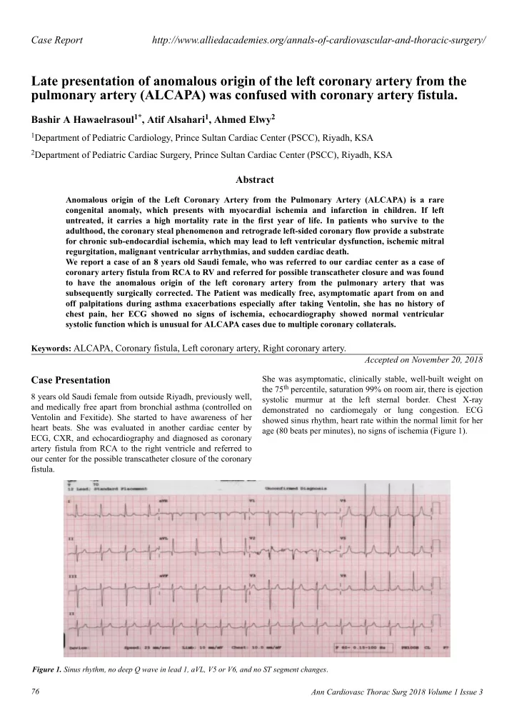
chest pain, her ECG showed no signs of ischemia, echocardiography showed normal ventricular systolic function which is unusual for ALCAPA cases due to multiple coronary collaterals. Keywords: ALCAPA, Coronary fistula, Left coronary artery, Right coronary artery. Accepted on November 20, 2018 Case Presentation She was asymptomatic, clinically stable, well-built weight on the 75 th percentile, saturation 99% on room air, there is ejection 8 years old Saudi female from outside Riyadh, previously well, systolic murmur at the left sternal border. Chest X-ray and medically free apart from bronchial asthma (controlled on demonstrated no cardiomegaly or lung congestion. ECG Ventolin and Fexitide). She started to have awareness of her showed sinus rhythm, heart rate within the normal limit for her heart beats. She was evaluated in another cardiac center by age (80 beats per minutes), no signs of ischemia (Figure 1). ECG, CXR, and echocardiography and diagnosed as coronary artery fistula from RCA to the right ventricle and referred to our center for the possible transcatheter closure of the coronary fistula. Figure 1. Sinus rhythm, no deep Q wave in lead 1, aVL, V5 or V6, and no ST segment changes . 76 Ann Cardiovasc Thorac Surg 2018 Volume 1 Issue 3

  2. Hawaelrasoul et al. 24 hour Holter monitoring showed normal sinus rhythm, maximum heart rate of 152, minimum heart rate was 53 and average heart rate was 79 beats per minutes and no documented ventricular or supraventricular ectopy. Echocardiography showed: large coronary fistula seen in MPA. LCA origin was not clarified. Mild left atrial enlargement. Moderately dilated left ventricle, there was trivial mitral regurgitation with good left ventricle systolic function. Some coronary collaterals saw in the anterior interventricular groove. Further imaging was recommended is to clarify coronary anatomy (Figures 2-5). The case was discussed in the cardiology meeting next day and there was a large debate about is it coronary fistula or ALCAPA. Figure 4. 3D-echo showing good LV function with no regional wall abnormality . Figure 2. 2D echocardiography and color Doppler confirm the origin of LCA from MPA . Figure 5. Longitudinal starin is -24.8%, EF 61.7%. Figure 3. M-mode showing Good LV systolic function EF 62%. Figure 6. Selective right coronary angiogram via right femoral artery illustrating the presence of large tortuous right coronary RCA. Next day the patient was taken to the catheter laboratory and selective coronary angiography showed dilated RCA with multiple collaterals supplying the LCA which drain retrogradely to the MPA (Figures 6 and 7). 77 Ann Cardiovasc Thorac Surg 2018 Volume 1 Issue 3

  3. Citation: Hawaelrasoul BA, Alsahari A, Elwy A. Late presentation of anomalous origin of the left coronary artery from the pulmonary artery (ALCAPA) was confused with coronary artery fistula. Ann Cardiovasc Thorac Surg 2018;1(3):76-80. known, with only a few case reports of patients older than 50 years of age [1]. The true incidence of older patients is not known [2,3]. It is unusual for an ALCAPA patient to survive to adulthood, however, the oldest reported patient with ALCAPA to undergo corrective surgery was a 79-year-old woman presented with a 3-months history of worsening shortness of breath and orthopnea [4]. Invasive coronary angiography is recognized as the “gold standard” for the diagnosis of coronary artery disease because of its excellent spatial and temporal resolution. Diagnostic cardiac catheter with coronary angiogram illustrated enlarged right coronary artery, which provides retrograde collaterals to supply the left ventricle then preferentially directs into the lower pressure pulmonary artery system causing coronary steal phenomenon. Few patients who survive through adulthood without surgery must have abundant, well-formed functioning collaterals with adequate perfusion of the left ventricle [5]. Multislice Computed Tomography (MSCT) is a non-invasive imaging tool that allows accurate, non-invasive diagnosis of Figure 7. Diagnostic coronary angiogram via right femoral artery ALCAPA, it has been increasingly used to assess the coronary illustrating the presence of a large tortuous right coronary artery anatomy. In recent years with very good diagnostic accuracy (RCA) which gives multiple collaterals filling the left coronary and is considered a reliable alternative to invasive coronary arterial system (LCA) and retrograde flow of contrast within the main angiography for diagnostic evaluation of the coronary anatomy pulmonary artery (PA). [6]. However, ECG gating of the scans requires heart rates to be relatively slow and there remains a degree of radiation Hemodynamics showed:- QP: QS was 1.5:1. Pulmonary exposure with this technique, which is difficult in children vascular resistance (PVR=1) wood unit, Cardiac Index (CI) 9.7 even with the assistance of B-blocker therapy as used in adult litres/minute/m 2 . Right Pulmonary artery pressure 19/12 patients. mmHg with a mean of 16 mmHg. LV end-diastolic pressure 10 mmHg and RV end-diastolic pressure 8 mmHg, systemic right Echocardiography with Doppler color flow mapping has femoral pressure 94/55 with a mean of 71 mmHg. replaced cardiac catheterization as the standard method of diagnosis [7]. An enlarged RCA should also raise the suspicion After confirmation of diagnosis, surgery was done on the same of the diagnosis. Echocardiography also will show the size and admission by coronary artery implantation through median function of the cardiac chambers, particularly the left ventricle, sternotomy pericardium opened after heparinization, the aorta as well as regional left ventricular wall motion abnormalities and double stage cannulation were done, bypass commenced and mitral regurgitation. There may be increased echogenicity and patient gradually cooled down to 28-degree centigrade. of the papillary muscle and adjacent endocardium due to Aorta was cross-clamped and heart cardioplegia, LV vent was fibroelastosis. inserted through a right upper pulmonary vein. MPA was transected near the confluence. LCA button harvested and The typical and direct echocardiographic feature of ALCAPA mobilized then re-implanted to the aorta, the aortic clamp was is the ostium of the LCA originating from the pulmonary artery removed after careful de-variation. The heart was beating in in parasternal short-axis view. It is difficult to distinguish LCA sinus rhythm, PA reconstructed with a pericardial patch. The originating from the pulmonary artery or the aorta based on patient came out of bypass easily on moderate inotropic anatomical image analysis, because the LCA is close to the support, the heart was decannulated and hemostasis was aorta, and sometimes it even crosses through the aortic wall in secured. The chest was closed with 2 pleuro-mediastinal patients with ALCAPA. The blood flow in the abnormal vessel drains. Intraoperative transesophageal echocardiography from the pulmonary trunk is in the opposite direction of normal showed patent coronary arteries with good LV systolic LCA and this is prominent in the diastolic phase. In some function. The patient was shifted to surgical ICU in stable cases, the two branches of LCA and retrograde flow can be condition with the good postoperative course. On clinical visualized. However, when visualizing a dilated RCA and follow-up, she is completely asymptomatic, last diastolic or continuous retrograde flow from the abnormal echocardiography showed normal left ventricle dimension, vessel into the main pulmonary artery, can be misdiagnosed as trivial mitral regurgitation, and good left ventricle function. RCA to pulmonary artery fistula [8]. In our patient she was completely asymptomatic, thriving well Discussion apart from dyspnea on moderate exertion and palpitation which ALCAPA is a very rare congenital cardiovascular defect with can be explained by uncontrolled bronchial asthma, which led an estimated incidence of one in 300,000 live births; however, her to seek medical advice. She has no chest pain or signs of this may be an underestimated considering patients may die prior to diagnosis. The true incidence of older patients is not 78 Ann Cardiovasc Thorac Surg 2018 Volume 1 Issue 3

Download Presentation
Download Policy: The content available on the website is offered to you 'AS IS' for your personal information and use only. It cannot be commercialized, licensed, or distributed on other websites without prior consent from the author. To download a presentation, simply click this link. If you encounter any difficulties during the download process, it's possible that the publisher has removed the file from their server.

Recommend


More recommend