Laser-hybrid accelerator for radiobiology (LhARA) Cancer and - - PowerPoint PPT Presentation

laser hybrid accelerator for radiobiology lhara
SMART_READER_LITE
LIVE PREVIEW

Laser-hybrid accelerator for radiobiology (LhARA) Cancer and - - PowerPoint PPT Presentation

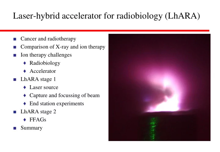
Laser-hybrid accelerator for radiobiology (LhARA) Cancer and radiotherapy Comparison of X-ray and ion therapy Ion therapy challenges Radiobiology Accelerator LhARA stage 1 Laser source Capture and focussing of beam


slide-1
SLIDE 1

Laser-hybrid accelerator for radiobiology (LhARA)

■ Cancer and radiotherapy ■ Comparison of X-ray and ion therapy ■ Ion therapy challenges ♦ Radiobiology ♦ Accelerator ■ LhARA stage 1 ♦ Laser source ♦ Capture and focussing of beam ♦ End station experiments ■ LhARA stage 2 ♦ FFAGs ■ Summary

slide-2
SLIDE 2

Cancer and radiotherapy

■ Globally, one in 5 men and one in 6 women develop cancer during their lifetime (WHO – 2018). ■ Radiotherapy indicated in about 50% of cases. ■ One in 8 men and one in 11 women die from the disease. ■ Provision of treatment on the necessary scale requires development of new techniques… ■ ….And will generate substantial economic impact. ■ Number of people served by one radiotherapy unit:

2

slide-3
SLIDE 3

Proton and ion therapy

■ Proton therapy first proposed in “Radiological Use of Fast Protons”, R.R. Wilson, Radiology 47:487- 491, 1946. ■ Robert Wilson was involved in the Manhattan Project, was a sculptor, architect and the first director of Fermilab. ■ First patients1954 at LBNL. ■ “Pituitary irradiation with high-energy proton beams: a preliminary report.” C.

  • A. Tobias et al. Cancer Res. 18(2):121-

34, 1958.

3

slide-4
SLIDE 4

An aside

■ When called to justify the cost of Fermilab to a congressional committee, Wilson said: ■ “It only has to do with the respect with which we regard one another, the dignity of men, our love of culture... It has to do with: Are we good painters, good sculptors, great poets? I mean all the things that we really venerate and honor in our country and are patriotic about. In that sense, this new knowledge has all to do with honor and country but it has nothing to do directly with defending our country except to help make it worth defending.”

4

slide-5
SLIDE 5

Proton and ion therapy

■ Existing proton and carbon beam treatment facilities: ■ In contrast, well over 10 000 linacs used for X-ray therapy. ■ Compact, (relatively) cheap, can be installed in most hospitals.

1 1 1 1 6 3 3 3 1 1 1 3 2 1 4

1

1 2 3

14 6

2 1

33

1

Carbon ion facility(13 total) Proton beam facility(83 total)

5

slide-6
SLIDE 6

X-ray and ion therapy

■ Ion therapy has advantages over X- rays due to energy loss mechanisms for photons and ions in material. ■ Get exponential dose depth profile for photons, “Bragg peak” for ions: ■ Clinical treatments use a Spread Out Bragg Peak (SOBP) to conform dose

  • ver the length of the tumour.

■ Carbon ions have a better dose profile in front of the tumour than protons, though fragmentation creates a distal tail.

Levin et al. “Proton beam therapy” British Journal of Cancer volume 93, pages 849–854 (2005).

6

slide-7
SLIDE 7

X-ray and ion therapy

■ Volumetric Modulated Arc Therapy (VMAT) therapy delivers X-ray dose conformal to tumour by modulating beam while rotating around patient. ■ Here for prostate cancer patient: ■ Minimises dose to surrounding tissues. ■ Compare with dose distribution from four-beam intensity modulated treatment plan using protons: ■ Allows reduced dose to critical tissues, here, bowel and spine.

7

slide-8
SLIDE 8

Proton therapy in the UK

■ Douglas Cyclotron at Clatterbridge produces 60 MeV protons. ■ Originally designed to provide fast neutron therapy for trials of radio- resistant tumour treatment. ■ Total of 384 patients treated with neutrons until 1995. ■ Now used solely for proton therapy

  • f eye tumours.

■ About 450 diagnosed in the UK each year, 40% of patients referred for proton-beam radiotherapy. ■ Age of eye patients from 9 to 92 years, average about 50 years. ■ Some beam time for research.

8

slide-9
SLIDE 9

Proton therapy in the UK

■ Two new NHS centres, each to treat about 750 patients per annum. ■ Use cyclotrons, producing 230 MeV proton beam (range 33 cm in water). ■ Carbon absorbers can reduce energy to 70 MeV. ■ One centre, Christie NHS Foundation Trust (Manchester), opened in 2018: ■ Second centre, University College London Hospital NHS Foundation Trust, due to open in 2020: ■ Also Rutherford Cancer Centre machines in Newport, Bedlington and Reading, and one under construction in Liverpool. ■ All 230 MeV cyclotrons.

9

slide-10
SLIDE 10

Ion therapy challenges – radiobiology

■ Biological effects of a given dose of radiation (energy deposited per kg, units Gy) depend on the type of radiation (and other factors). ■ Radiobiological effectiveness (RBE) is ratio of effectiveness w.r.t. photons. ■ Typically, constant RBE = 1.1 used for planning proton therapy. ■ For carbon ions, RBE typically above about 2.25. ■ Systematic studies needed to improve data and understanding of underlying radiation damage mechanisms. ■ Important for planning of proton and ion treatments.

Experimental proton RBE values relative to 60Co. Paganetti H et al. 2002 Relative biological effectiveness (RBE) values for proton beam therapy

  • Int. J. Radiat. Oncol. Biol. Phys. 53 407–21

10

slide-11
SLIDE 11

Ion therapy challenges – radiobiology

■ Need for better understanding illustrated by efficacy of Ultrahigh Dose-rate (FLASH) radiotherapy. ■ Conventional treatment, dose rate typically a few Gy/min. ■ Flash treatments, dose rates above 100 Gy/s. ■ Efficacy higher for a given dose. ■ Evidence that normal tissue sparing is also improved, e.g. “The Advantage

  • f FLASH Radiotherapy Confirmed

in Mini-pig and Cat-cancer Patients.” Vozenin et al. Clin. Cancer Res. 2019 Jan 1;25(1):35-42. ■ Microbeam radiation therapy showing similarly interesting results.

Thirty-six weeks postradiotherapy, quantification of hair follicles per tissue section performed on sections cut from 6-mm punch biopsies taken from nonirradiated, Conv-RT, and FLASH-RT patches (data are presented as number of hair follicles per tissue section).

11

slide-12
SLIDE 12

Ion therapy challenges – accelerator

■ Cyclotrons deliver beams at final, single energy. ■ Degraders used to decrease beam energy, cause scattering, produce radiation. ■ Low energy injection of cyclotrons limits beam current due to space charge. ■ Cyclotrons one ion species. ■ Synchrotrons can accelerate multiple ion species but larger and more costly. ■ Gantries, significant cost of the accelerator facility. ■ MedAustron p and C synchrotron: ■ Varian proton gantry:

12

slide-13
SLIDE 13

LhARA

■ Radiobiology research facility. ■ Initial acceleration laser driven. ■ Gives large flux of protons and ions in very short bunches. ■ Compact capture system. ■ Injection at 15 MeV overcomes space charge limitations. ■ Allows large instantaneous doses. ■ Implementation in two stages. ♦ In vitro studies, 15 MeV protons. ♦ In vivo studies, 127 MeV protons, in vitro studies with 33 MeV/u carbon 6+ ions. ■ Groups involved:

13

slide-14
SLIDE 14

LASER TARGET Laser used to generate intense beams of different types of ions, e.g. protons and carbon ions. CAPTURE SECTION Gabor lenses used for compact focussing to capture the large divergence and energy spread of the laser-driven ion beam. ENERGY SELECTION A Gabor lens and a collimator are used to select particle energy.

z y

45° 11.58 m

90° VERTICAL BEND Combined function magnets deliver the beam vertically to the end station. MATCHING Further Gabor lenses are used to adjust the beam size and divergence in the end station. END STATION Where the cells are

  • irradiated. The beam is

delivered vertically from below the cell culture plate.

beam

2.55 m

LhARA stage 1

■ Facility for irradiating cell samples.

14

slide-15
SLIDE 15

Laser source

■ Target normal sheath acceleration mechanism. ■ Protons from (hydrocarbon) contamination on foil surface. ■ Produces large flux of ions in pulse

  • f ~ 30 fs duration.

■ Kinetic energy up to 15 MeV.

Laser driven ion beam simulation using EPOCH. Energy vs angle with respect to laser beam direction

15

slide-16
SLIDE 16

Capture and focussing of beam from source

■ Initial beam has small size, large divergence, large energy spread. ■ Capture and focussing difficult. ■ Approaches include magnetic systems based around: ♦ Quadrupole magnets. ♦ SC solenoidal magnet. ♦ Pulsed solenoidal magnet. ■ Here, propose to use Gabor lenses. ■ Principle of Gabor lens: ■ Electron cloud confined axially by anode (red) and grounded electrodes (black). ■ Radial confinement produced by magnetic field. ■ Space charge due to electron cloud focusses protons. ■ RA ~ 20 mm, RC ~ 10 mm, L ~ 0.5 m. ■ VA ~ 600 kV, BZ ~ 0.3 T.

16

slide-17
SLIDE 17

Capture and focussing of beam from source

■ Prototype Gabor lens constructed at Imperial College: ■ Initial measurements with prototype using 1 MeV proton beam at Surrey Ion Beam Centre. ■ Gabor lens design being updated at Imperial. ■ Next steps: ■ Vacuum tests. ■ Tests with radioactive source. ■ Tests with the laser driven ion beam.

17

slide-18
SLIDE 18

Capture and focussing of beam from source

■ Example of section of Gabor lens lattice: ■ Lens 1: Capture large fraction of particles, reduce divergence angle. ■ Lens 2: Focus beam into aperture for energy separation. ■ Lens 3: Deliver beam to lenses and magnets of end-station section.

18

slide-19
SLIDE 19

Beam transport

■ Simulate beam transport from source through to target station using BDSIM (Beam Delivery Simulation), based on Geant4. ♦ Model beam line elements. ♦ Track through 3D EM fields. ♦ Simulate material interactions. ♦ Extract beam optics parameters and energy deposition. ■ While design evolving, Gabor lenses modelled as equivalent focal length solenoidal magnets. ■ Shift to EM simulation of lenses at later stage. ■ Initial beam generated by BDSIM. ■ Tracked through LhARA:

19

slide-20
SLIDE 20

End station

■ Beam energy chosen to ensure Bragg peak can be scanned through cell layer. ■ Detailed simulation of delivery of beam to sample.

20

10 MeV 12 MeV 15 MeV

0.075 mm window

beam

0.03 mm cells 1.15 mm base 5 mm air gap 0.25 mm scint. fibre layer 15 mm cell nutrients

Energy loss as function of depth

slide-21
SLIDE 21

Example of end-station experiments

■ Understand damage caused to DNA by proton irradiation. ■ Single strand breaks rapidly repaired. ■ Repair of double strand breaks more difficult. ■ Study number of single and double strand breaks as a function of dose, dose rate… ■ Determine damage e.g. using Comet analysis: ♦ Irradiate cells and embed in agarose gel. ♦ Forms mesh through which fluids can permeate, but traps cells. ♦ Perform lysis to break down cell membrane and membrane surrounding nucleus. ♦ Leaves DNA in holes originally

  • ccupied by cells.

21

slide-22
SLIDE 22

Example of end-station experiments

■ Comet analysis cont. ♦ Electrophoresis causes charged DNA to migrate through mesh. ♦ Speed of migration depends on length of DNA strands. ♦ Stain and image the “comets”. ■ Comet analysis cont. ♦ Amount of light from tail contains information about number of DNA strand breaks. ♦ Compare with image of similarly processed, unirradiated cells:

22 Jason Parsons, Liverool Univ.

slide-23
SLIDE 23

LhARA – stage 2

■ Goal – irradiate animal models. ■ Need post-acceleration to increase energy of the beam driven by the laser. ■ Current solution is based on a fixed field alternating gradient accelerator. ■ Can achieve factor 3 or more increase in momentum. ♦ Accelerate 15 MeV protons to 127 MeV. ♦ 3.8 MeV/u carbon 6+ ions to 33 MeV/u.

23

slide-24
SLIDE 24

Fixed field alternating gradient accelerators

■ FFAGs are accelerators in which magnetic fields do not vary in time but are a strong function of radius. ■ Orbit changes with momentum are relatively small. ■ Radius of orbit given by ■ Scaling FFAG, orbit shape independent of momentum. ■ Non-scaling FFAG, shape of orbit is function of momentum. ■ Scaling FFAG, k ~3. ■ Non-scaling FFAG, k can be as large as 80.

( )

1

.

k A

r r p p =

Low energy High energy

D D F

Low energy High energy

D D F

Beam dynamics (and orbits) more complex than in cyclotron. 24

slide-25
SLIDE 25

FFAGs

■ Proposed for use as muon accelerators, in accelerator driven sub-critical reactors, and for medical applications. ■ Advantages over conventional medical cyclotrons. ♦ Variable energy operation without energy degraders. ♦ Operation with different ions. ♦ Multiple extraction ports. ■ FFAG concept developed independently in 1950s in Japan by Tihiro Ohkawa, in the US by Keith Symon, and in Russia by Andrei Kolomensky. ■ Initial designs all scaling FFAGs. ■ First 500 keV e‒ prototype operated in Michigan 1956.

25

slide-26
SLIDE 26

FFAGs

■ First 500 keV p FFAG, KEK in 2000. ■ FFAG for ADSR, Kyoto University ■ First non-scaling FFAG, EMMA, at Daresbury. ■ Acceleration of electrons from 10 to 20 MeV reported in Nature in January 2012.

26

slide-27
SLIDE 27

LhARA FFAG

■ LhARA ring: ■ LhARA ring parameters: ■ Number of sectors 10 ■ k 5.19 ■ Rmin 2.92 m ■ Rmax 3.49 m ■ Bmax 1.4 T ■ Max p injection E 15 MeV ■ Max p extraction E 127 MeV ■ RF freq 2.89…6.48 MHz ■ Bunch intensity 108 protons

27

slide-28
SLIDE 28

LhARA magnets and RF

■ Spiral FFA magnet ■ Built by SigmaPhi for the RACCAM project. ■ Need cavities capable of low frequency and large frequency variation. ■ Can use magnetic alloy (MA) cavities, similar to those used at J-PARC.

28

slide-29
SLIDE 29

Induction acceleration for FFAGs

■ Circumvent difficulties of synchronising RF in an FFAG by using “betatron” acceleration mechanism? ■ Faraday’s law: ■ Varying magnetic flux through centre of FFAG generates EMF which accelerates particles. ■ Design of an electron FFAG for industrial applications was studied in the USA by RadiaBeam Technologies. ■ Radiatron: ■ Reported in Proc.s of PAC07 (Albuquerque) and of EPAC08 (Genoa)…but then development stopped.

2

.

A

r

d E ds dt

 = = −

29

slide-30
SLIDE 30

Induction acceleration for LhARA

■ Look at possibility of using induction acceleration for LhARA. ■ Set up LCR circuit by adding C to L

  • f accelerating magnets.

■ Choose C to get appropriate resonant frequency. ■ Resulting acceleration of proton beam injected at 15 MeV: ■ May be worth looking at a little more seriously. EMF (V)

1 10 3

 2 10 3

 3 10 3

 30 − 20 − 10 − 10 20 30 4 − 105  2 − 105  2 105  4 105  2 2 −  .B

( )

 − 

( )

( )

( )

( )

    +

( )

     

Flux (W) t (s)

6 10 4

 8 10 4

 1 10 3

 1.2 10 3

 1.4 10 3

 5 107  1 108  1.5 108  0.01 0.02  

( ) ( ) ( ) ( )

( )

 +

( ) ( ) ( )

( )

 +

( ) ( )

( )

3 −

4 −

 t0r t1r  t2r  t1r 

t (s) Energy spread Energy (eV)

30

slide-31
SLIDE 31

FFAG gantry

■ Reduce gantry cost and mass by using FFAG. ■ E.g. of design for PAMELA. ■ Produces zero dispersion condition at end of first bending section and at end of gantry.

31

slide-32
SLIDE 32

Summary

■ Cancer is a major threat to health. ■ Radiotherapy is a valuable treatment method in about 50% of cancer cases. ■ Proton/ion therapy offers significant advantages over X-rays. ■ Potential large benefits through FLASH treatment. ■ But further radiobiology studies needed. ■ Wider provision of ion therapy requires significant cost reductions. ■ LhARA is attempt to couple development of technologies for proton/ion therapy with radiobiology experiments. ■ Ultimate goal (2050?), thorough understanding, optimisation of radiobiology of proton/ion therapy. ■ Low cost proton/ion therapy system based on laser acceleration and Gabor lenses for beamline/gantry.

32