SLIDE 2 2
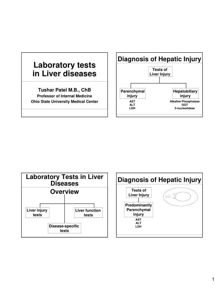
Predominantly Parenchymal injury Tests of Liver Injury
AST ALT LDH
Diagnosis of Hepatic Injury
Enzymes leak out
Alanine Aminotransferase (ALT) Glutamate Pyruvate Transaminase
- As a group, the aminotransferases catalyze the
interconversion of amino acids and alpha-keto acids by transfer of amino groups.
- Highest activity in the liver, with decreasing
concentrations in the kidney, heart, skeletal muscle, pancreas, spleen, and lung.
- The enzyme is mainly located in the cytoplasm
and escapes into the blood circulation during the cellular injury.
Aspartate aminotransferase (AST) Glutamate-Oxaloacetate Transaminase
- Ubiquitous, and found in all tissues
- Highest activity in heart, with significant activity
in the liver, brain, gastric mucosa, adipose tissue, skeletal muscle, and kidneys.
- In mild hepatocellular injury when the plasma
but not the mitochondrial membrane is damaged, cytoplasmic form of AST is released into the blood. With more severe hepatocellular injury, mitochondrial damage may result in the release of mitochondrial AST.
- CPK or aldolase may be useful to exclude a
muscle source of abnormal AST
Increased AST/ALT
- Increased ALT or AST usually imply hepatic
parenchymal injury but can occur in other organ damage ALT is more “liver” specific.
- Reflect hepatic injury not hepatic function – and
can be normal in advanced liver disease or cirrhosis
- Can occur with damage to other organs
(myocardial infarction, muscular dystrophy, etc)