SLIDE 7 8/16/2016 7
NOVEL ALGORITHM OF DOMINANT FREQUENCY & ELECTROGRAM PATTERNS TO IDENTIFY FOCAL SOURCES AND ROTATIONAL PATTERNS DURING HUMAN PERSISTENT ATRIAL FIBRILLATION
AtulVerma, MD, FHRS, Thomas Deneke, MD, PhD, Yariv A. Amos, Msc, Roy Urman, BSc, Philipp Halbfass, MD, Karin M. Netwich, MD, Erik Wissner, MD, FHRS, Karl- Heinz Kuck, MD, FHRS and Roland, TilzMD.
SOUTHLAKE REGIONAL HEALTH CENTRE, Newmarket, Ontario, Canada; HEART CENTER BAD NEUSTADT, Bad Neustadt, Germany; BIOSENSE WEBSTER, Haifa, Israel; ASKLEPIOS KLINIK ST. GEORG, Hamburg, Germany
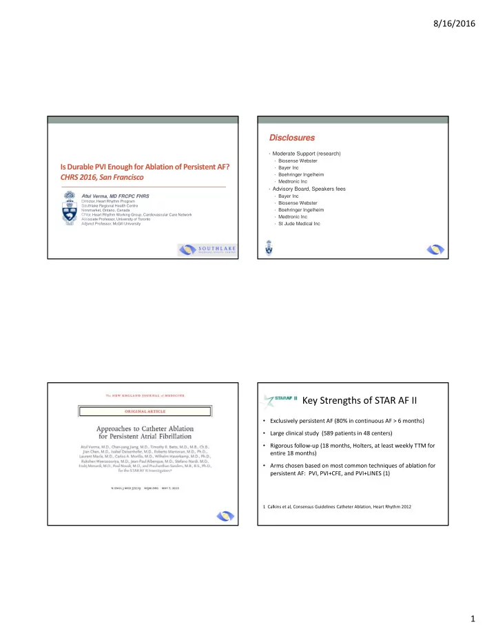
Automated methods may identify areas of interest during ablation of persistent atrial fibrillation (AF). We sought to determine if an algorithm based on dominant frequency (DF) and electrogram (EGM) patterns during persistent AF could be used to identify focal sources and rotational patterns. Maps of persistent AF were acquired using a multi-electrode basket catheter. The CARTOFINDER™ algorithm filters all unipolar EGMs for quality, reduces far-field ventricular artifacts and annotates the timing
- f the activation wave front. DF analysis was
performed and the pattern was classified as homogenous (<0.5 Hz) or heterogeneous (>0.5 Hz) & stable (regular/no variation over 30 sec) or unstable (random variation). The algorithm can identify QS EGM patterns and “regular” sequential atrial activation gradients occupying >50% of the cycle length (CL) suggesting rotational wave
- fronts. All 4D activation maps were reviewed
by two blinded independent adjudicators to visually identify focal sources and rotational wave fronts & only those agreed upon by both reviewers were included for analysis. A CARTOFINDER™ algorithm based on DF, LAT and EGM patterns correlated well with visually confirmed regions of interest during human persistent AF. These patterns could prospectively identify regions of interest with a reasonably high predictive value. Rotational activations covered 67±8% of the local CL with a mean of 3.0±2.7 rotations. Rotational wave front patterns were related to areas of homogeneous-stable DF spatiotemporal stability (variance 0.46± 0.17Hz) and with sequential EGM activation gradients . Focal sources were related to areas of heterogeneous-stable DF spatiotemporal stability (variance 0.58±0.30Hz) and with a consistent QS EGM
- pattern. DF temporal stability between RAPs
and sources was statistically significantly different (unpaired t test, p<0.000001)
CARTOFINDER™ Algorithm sensitivity and specificity with respect to human expert identification Foci Sensitivity/Specificity 89% / 77% RAP Sensitivity / Specificity 82% / 70%
ABSTRACT METHODS The CARTOFINDER™ system identified 34±14% of the basket EGMs were adequate for analysis when positioned in the left atrium (LA) and 60±15% were adequate in the right atrium (RA). 20 patients were analyzed, rotational activations were identified by the experts in 27/70 (39%) of the LA maps and 24/51 (47%)
- f the RA maps. Focal sources were
identified in 47/70 (67%) of the LA maps and 46/51 (90%) of the RA maps. . RESULTS Focal Source Rotational Activation Pattern CONCLUSIONS
Number of focal sources found in analysis of 121 maps Number of rotational activation wave front found in analysis of 121 maps
TOUCH AF Trial – Verma et al, pending
- Contact force sensing for ablation of persistent AF
- Allowed wide antral PVI and roof line only
- Randomized operators to CF-guided vs CF-blinded
approach
- Final analysis not yet complete, BUT…..
- Average contact force was 15 grams for both arms
- Overall success rate was 78% with fewer than 20% of
patients requiring second procedure
26
Conclusions
- PVI seems to be the cornerstone for any ablation
procedure for persistent AF
- Success rates with PVI alone seem to be stuck around
60%
- Many will require more than one procedure
- Mapping AF may help us to realize novel targets for
ablation and improve success rates