
SLIDE 1
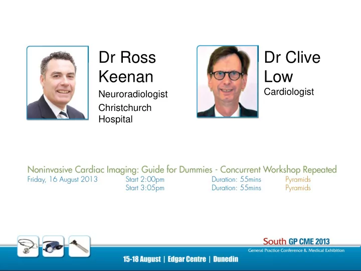
Dr Ross Keenan
Neuroradiologist Christchurch Hospital
Dr Clive Low
Cardiologist
Keenan Low Cardiologist Neuroradiologist Christchurch Hospital - - PowerPoint PPT Presentation
Dr Ross Dr Clive Keenan Low Cardiologist Neuroradiologist Christchurch Hospital Heart Vision Non Invasive Cardiac Imaging A Guide for Dummies Drs Clive Low (Cardiology) & Ross Keenan (Radiology) GP CME, Dunedin 16 August 2013
Dr Ross Keenan
Neuroradiologist Christchurch Hospital
Dr Clive Low
Cardiologist
Dr R J Keenan CRG 2007
“Non Invasive Cardiac Imaging – A Guide for Dummies” Drs Clive Low (Cardiology) & Ross Keenan (Radiology) GP CME, Dunedin 16 August 2013 Workshop, Friday 2-4pm Edgar Centre
R J Keenan CRG 2010 joint venture
CHRISTCHURCH RADIOLOGY GROUP & HEART CENTRE (2003) www.heartvision.co.nz
R J Keenan CRG 2012 Non Invasive Cardiac Imaging
A Guide for Dummies Dr Clive JS Low Consultant Cardiologist
CT Coronary Calcium Score
An in-patient susceptibility study
The validation studies in Males aged 50 to 70 show us that little or no calcified plaque identifies low risk of IHD events in the patient Studies demonstrate more accurate risk prediction For an individual patient by combining the Framingham Risk (NHF table) and CT calcium score
CT Coronary Calcium Score
An in-patient susceptibility study
A patient with high calcium score (≥ 300 Agaston units) has the same IHD event rate as a patient who has had MI, CABG, PTCA, or abnormal coronary angiogram
CT Coronary Angiogram
An accurate test for diagnosis of IHD
Ex ECG 75% accurate Stress Echo (and all the others) 80ish% accurate For Obstructive IHD CT Coronary Angiogram 98% accurate For Significant coronary atheroma (NB All quite operator dependant)
CT Coronary Angiogram
An accurate test for diagnosis of IHD
Limitations Radiation exposure esp young and females Contrast exposure allergy Arrhythmia ectopic beats atrial fibrillation tachycardia (NB ?β Blocker) Severe disease high risk patients calcium bloom
www.heartvision.co.nz
R J Keenan CRG 2012 Right to Left: Amanda, Jo, Dr Latham Berry, Dr Sharyn Macdonald, Dr Ross Keenan, Jenny, Clare, Rachel
Dr R J Keenan CRG 2012
Cardiac Imaging Team
Dr R J Keenan CRG 2007
Cardiac CT : Heart Vision
CACS “screening” not covered CCTA - SXHI criteria v others
R J Keenan CRG 2012 Siemens Dual Source CT: Left: Definition 2007 (St Georges), Right: Definition FLASH 2012 (Christchurch Hospital)
Cardiac CT Imaging Systems
Heart Vision : Dr Sharyn Macdonald, Cardiac Radiologist: Siemens Dual Source Definition CT_Circulation analysis
Cardiac CT Imaging Systems
Left: CCTA Syngo via VR Right: Syngo via curved MIP, normal LAD
Cardiac CT Imaging Systems
Dr R J Keenan CRG 2007
‘5’ learning points
Dr R J Keenan CRG 2007
Dr R J Keenan CRG 2013
Cardiac CT System
Siemens Dual Source CT
Dr R J Keenan CRG 2007
CCTA Radiation Dose - 2009
Technique Effective Dose pa SPECT Thallium stress 25mSv SPECT Sestamibi stress 12-18mSv CT chest ungated helical 5-7mSv Retrospective mode CCTA 14 mSv, (4.5-19) < 5-6mSv Prospective mode CCTA 5 mSv,(1.7-7.3) < 3-4mSv FLASH mode CCTA </= 1mSv Diagnostic catheter angiogram 3-6 mSv, (3-30) CXR (PA/Lat) 0.05 mSv Annual background radiation 2-5mSv (~ 3mSv) Additional background radiation at altitude + 1.5mSv USA East-West round trip flight + 0.03mSv
Reference: Stolzmann P et al. Eur Radiol 2008; 18: 592-599
R J Keenan CRG 2009 Dose
CCTA Radiation Dose - 2009
0.0 5.0 10.0 15.0 20.0 25.0 30.0 35.0 40.0 45.0 50.0 50 100 150 200 250 300
CT upgrade 2009 Prospective Adaptive Sequence Retrospective “min dose 4%” CT upgrade 2012 Prospective min dose Adaptive Sequence FLASH Cardio mode IR - SAFIRE CT 2007 Retrospective Spiral
Dr R J Keenan CRG 2007
CCTA Radiation Dose - 2012
R J Keenan CRG 200 R J Keenan CRGTechnique Mean Dose (mSv) Retrospective gated < 6mSv Prospective gated 0.8 - 4mSv Siemens FLASH mode << 2mSv CACS < 0.5mSv
Reference: Heart Vision Audit 2011:
R J Keenan CRG 2012 Dr R J Keenan CRG 2007
Coronary Artery Calcium Scoring
R J Keenan CRG 2010 CACS
R J Keenan CRG 2010Non-contrast CACS Contrast CCTA
Dr R J Keenan CRG 2007
CACS
Framingham CAD Risk Profile Low risk < 10% /10 year risk cardiac event → CCTA Intermediate risk ~10-20% /10 year risk cardiac event → CCTA High risk > 20% /10 year risk cardiac event → DSA
MISSES (?10-25%)
CAD Risk Stratification: definitions
CVD Risk Stratification
Event Free Survival Follow-up 1.7% Normal 2.7% 1V NOD 4.6% 2V NOD 6.9% 3V NOD 7.1% 1V OD 11.3% 2V OD 20% 3V OD
NOD = non obstructive disease OD = obstructive disease
CACS CCTA
CACS - Interpretation
R J Keenan CRG 2012CACS Score (Agatston) Plaque burden Obstructive CAD Risk CVD Risk Guidelines none < 5% very low
1-10 minimal < 10% low
10-100 mild mild stenoses moderate
100-400 moderate NOCAD highly likely moderately high
> 400 severe > 90% risk of OCAD >/= 1 stenosis high
modification
Reference: Rumberger 1999
Dr R J Keenan CRG 2007
CACS
Dr R J Keenan CRG 2007
Coronary CT Angiography
R J Keenan CRG 2010 Dr R J Keenan CRG 2007
CCTA - Techniques
R J Keenan CRG 2009Retrospective Gating
Prospective Gating
FLASH Scan
CCTA Technique
Dr R J Keenan CRG 2012
ECG Pulsing
Dr R J Keenan CRG 2007
CCTA - Reporting Triage
Stenosis Grade:
borderline ~ 50% “significant” stenosis > 50%
“severe” stenosis > 70%
CCTA - Indications 1 Major
Reference: CSANZ November 2010
CCTA
R J Keenan CRG 2009 CCTA
M36yr Atypical CP. No Framingham risk factors. NETT. LAD > 90%
CCTA
R J Keenan CRG 2010FPH6911: M56yr ICU. Ex-smoker. Assess suitability as cardiac donor.
CCTA
R J Keenan CRG 2012LAD 50-60% LAD 50-60% DNC3450: M68yr CP. BETT. LBBB MR stress test -ve Rx medical
CCTA
R J Keenan CRG 2012LAD >70% LAD >70% LPG8917: M74yr Previous MVR. CT. BETT catheter + PCI
CCTA
R J Keenan CRG 2012LAD >70% LAD >70% AYD4723: M62yr CT. BETT. AF. FHx IHD catheter
CCTA “low-medium risk” (n=932) CAD (69%) Significant CAD (20%) Severe CAD (6%) Mild CAD (21%) CCTA normal (31%)
R J Keenan CRG 200 R J Keenan CRGHV Audit (2) 2008 - 2011
Reference: CCTA report analysis, HV Audit 2, Paula England June 2008 – August 2011 (n = 1002)
CCTA reported findings
CCTA v Catheter Concordance - Audit (2)
**discordant stenosis grade ≠ missed lesion
Dr R J Keenan CRG 2007
“Negative” CCTA - Prognosis
CVD events over the longer term (5yrs)
R J Keenan CRG 2010 Dr R J Keenan CRG 2007
Case Examples
R J Keenan CRG 2013Normal CACS & CTCA
CACS 98th centile, CTCA severe plaque
CACS 96th centile, CTCA moderate-severe
Normal CACS & CTCA
Dr R J Keenan CRG 2007
*SR*Intermediate Risk and SOB (known asthma)
Intermediate Risk and SOB
– Limited by dyspnoea – Moderately reduced Ex capacity – Borderline ST changes
Intermediate Risk and SOB
Case 1
R J Keenan CRG 2013CXR 2006 CXR 2011
Case 1
R J Keenan CRG 2013CT 2011
Case 1
R J Keenan CRG 2013CT 2011
Case 1
R J Keenan CRG 2013CT 2011
Case 1
R J Keenan CRG 2013CT 2011
Case 1
R J Keenan CRG 2013CXR 2011 preop CXR 2011 post lung transplant
Dr R J Keenan CRG 2007
Case 1
R J Keenan CRG 2013∆. Transbronchial biopsy = sarcoidosis. ∆. Open lung bx = UIP Rx → lung transplantation
Dr R J Keenan CRG 2007
*KN*Strong FHx severe IHD
predicted
*KN*Strong FHx severe IHD
Case 2
R J Keenan CRG 2013 Case 2
R J Keenan CRG 2013 Case 2
R J Keenan CRG 2013 *KN*Strong FHx severe IHD
– 30% 5yr risk !! – Risk is the same as previous IHD/2ͦ prevention
*KN*Strong FHx severe IHD
– If coronary calcification for on CT chest, or vascular calcification demonstrated during imaging for other causes (eg posterior tibial artery in ankle xray) patient may be at high coronary risk (>30%/5yr) and should be screened
Dr R J Keenan CRG 2007
Case 2
R J Keenan CRG 2013 Dr R J Keenan CRG 2007
*DD* Treated LDL, knee pain and SOB
2.6
– 3xCABG, 32(!!) Angiogram/plasties – 4 uncles CABG
*DD* Treated LDL, knee pain and SOB
injury 6/12 ago. Unable to run on treadmill @ gym.
*DD* Treated LDL, knee pain and SOB
Case 3
R J Keenan CRG 2013 Case 3
R J Keenan CRG 2013 Case 3
R J Keenan CRG 2013 Case 3
R J Keenan CRG 2013 Dr R J Keenan CRG 2007
Case 3
R J Keenan CRG 2013CABGs
catheter
Dr R J Keenan CRG 2007
*ES* Treated LDL, knee pain and SOB
*ES* Treated LDL, knee pain and SOB
typical angina 1mm inferolateral ST depression Stress Echo typical angina no wall motion defect
*ES* Treated LDL, knee pain and SOB
Case 4
R J Keenan CRG 2013 Case 4
R J Keenan CRG 2013 Dr R J Keenan CRG 2007
Case 4
R J Keenan CRG 2013 Dr R J Keenan CRG 2007
“Non Invasive Cardiac Imaging – A Guide for Dummies” Drs Clive Low (Cardiology) & Ross Keenan (Radiology)
R J Keenan CRG 2010