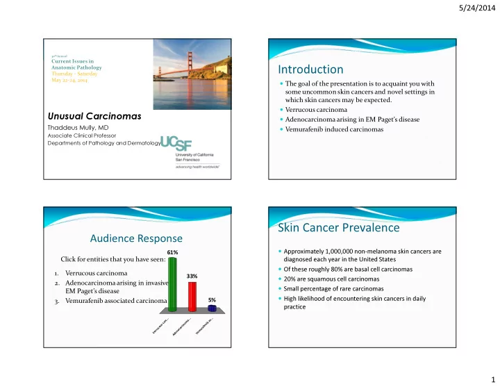
Introduction Thursday - Saturday May 22-24, 2014 The goal of the - - PowerPoint PPT Presentation

introduction
SMART_READER_LITE
LIVE PREVIEW

Introduction Thursday - Saturday May 22-24, 2014 The goal of the - - PowerPoint PPT Presentation

5/24/2014 30 th Annual Current Issues in Anatomic Pathology Introduction Thursday - Saturday May 22-24, 2014 The goal of the presentation is to acquaint you with some uncommon skin cancers and novel settings in which skin cancers may be


slide-1
SLIDE 1

5/24/2014 1

30th Annual

Current Issues in Anatomic Pathology Thursday - Saturday May 22-24, 2014

Unusual Carcinomas

Thaddeus Mully, MD

Associate Clinical Professor Departments of Pathology and Dermatology

Introduction

The goal of the presentation is to acquaint you with

some uncommon skin cancers and novel settings in which skin cancers may be expected.

Verrucous carcinoma Adenocarcinoma arising in EM Paget’s disease Vemurafenib induced carcinomas

Audience Response

Click for entities that you have seen:

V e r r u c

  • u

s c a r c . . . A d e n

  • c

a r c i n

  • m

a . . . V e m u r a f e n i b a s . . .

61% 5% 33%

1. Verrucous carcinoma 2. Adenocarcinoma arising in invasive EM Paget’s disease 3. Vemurafenib associated carcinoma

Skin Cancer Prevalence

Approximately 1,000,000 non-melanoma skin cancers are

diagnosed each year in the United States

Of these roughly 80% are basal cell carcinomas 20% are squamous cell carcinomas Small percentage of rare carcinomas High likelihood of encountering skin cancers in daily

practice

slide-2
SLIDE 2

5/24/2014 2

Verrucous carcinoma

Rare variant of squamous cell carcinoma First described in the oral cavity by Dr. L. Ackerman in

1948

Clinically characterized as a slowly growing verrucous

plaque at a variety of body sites (oral, genital, extremities)

May be locally destructive but seldom metastasizes

Verrucous carcinoma

Treatment is mainly surgical Large size of tumor and anatomic considerations lead

to propensity for local recurrence

May occasionally give rise to a high grade invasive

squamous cell carcinoma, especially in large lesions of long standing

Role of radiation is controversial

Photos courtesy of Dr. L. Requena

slide-3
SLIDE 3

5/24/2014 3

Verrucous carcinoma histopath

Usually a large sessile lesion Characterized by an exo-endophytic growth pattern “Pushing” rather than infiltrative margins “Deceptively” bland cytology Glassy eosinophilic keratinocytes Low mitotic activity Ample parakeratosis

slide-4
SLIDE 4

5/24/2014 4

slide-5
SLIDE 5

5/24/2014 5

Immunohistochemistry

Keratin is positive p53 usually noted just along basal layer, unlike more

conventional SCC which shows more haphazard changes

Ki-67 low and also at basal layer p16 typically negative

slide-6
SLIDE 6

5/24/2014 6

Ki-67 p16 p53 p53

slide-7
SLIDE 7

5/24/2014 7

p53

Human Papilloma virus

Is seldom found in association with verrucous

carcinoma

When papilloma virus is present transcriptionally

activated mRNA is not indicating that there is likely no causal role of papilloma virus in the generation of this type of carcinoma

Some have advocated use of papilloma virus testing to

aid in the differential diagnosis of this lesion

Pitfalls

Small biopsies may not demonstate all of the features

and lead to a benign diagnosis

Bland cytomorphology may lead to misdiagnosis as a

benign lesion

Summary

Large slowly growing tumor at a variety of body sites Characteristic architecture Bland cytomorphology Clinico-pathologic correlation important Small biopsies may be misleading

slide-8
SLIDE 8

5/24/2014 8

Adenocarcinoma in EMPaget’s disease

Extramammary Paget’s disease is a rare tumor of uncertain

  • rigin

Has been a “wastebasket term” and “primary” and “secondary” forms exist Primary thought to arise from either apocrine glandular or a stem cell in the epidermis Secondary indicates cutaneous involvement by an underlying visceral carcinoma (colon, bladder, prostate) Arises typically in apocrine rich areas including groin, perianal and scrotal areas Invasion is rare and portends a poor prognosis

Adenocarcinoma in EMPD

Typically a disease of older individuals (60’s-) Treatment is primarily surgical (with high recurrence

rates)

Rare occurrence in EMPD(<10%) but exact figures

hard to ascertain

Some advocate measurement of invasive component as

tumors > 1 mm in depth have worse prognosis with a greater propensity for lymph node and distant metastasis

Adenocarcinoma in EMPaget’s disease

Invasive cases may spread to regional lymph nodes and

metastasize to distant sites

Her-2-neu amplification has been shown to portend a

greater propensity for invasion, persistence, and nodal metastasis

This may be also be a therapeutic target

Photos courtesy of Dr. L. Requena

slide-9
SLIDE 9

5/24/2014 9

Adenocarcinoma histopathology

Typical intraepidermal changesof EMPD present As Paget’s may involve a very broad areas and result in large excisions careful scrutiny is necessary Immunohistochemistry is helpful in the differential diagnosis and potentially helpful in identifying small invasive foci, but beware in that normal glandular elements can be confounding One study demonstrated higher Ki-67 labelling rates and expression of cyclin D-1 in invasive versus non-invasive EMPD Serum Carcinoembryonic antigen (CEA) levels have also been shown to be elevated in invasive disease

slide-10
SLIDE 10

5/24/2014 10

Cytokeratin 7

slide-11
SLIDE 11

5/24/2014 11

slide-12
SLIDE 12

5/24/2014 12

Courtesy of Dr. L. Requena

Summary

Rare complication of Extramammary Paget’s Sampling important Thickness of lesion portends prognosis

slide-13
SLIDE 13

5/24/2014 13

Vemurafenib

Novel anti BRAF treatment Used in the treatment of metastatic melanoma that

harbors a BRAF V600E mutation

Shown to increase survival time in patients with

metastatic melanoma that harbors this mutation

A variety of cutaneous side effects have been described

both benign and malignant

Vemurafenib

39-year-old woman with a 7 year history of an irritated

lesion on the plantar left foot previously diagnosed as a plantar wart

Four month history of an enlarging subcutaneous

mass on the left calf

4X

Courtesy of A. Naujokas

Ulcerated T4b primary melanoma

Melan-A 10X Ki-67 20X

Staging workup

Primary Lesion: 10 mm thick ulcerated acral

melanoma primary.

Left calf: nodal melanoma metastases. Multiple other chest wall lesions noted, fine needle

aspiration of a right chest lesion consistent with metastatic melanoma.

Testing revealed a BRAF V600E mutation.

slide-14
SLIDE 14

5/24/2014 14

Stage IV BRAF mutant melanoma

Vemurafenib is a BRAF inhibitor that confers objective

tumor responses in half of patients treated.

Patient opted to initiate Vemurafenib

Overall Survival (6 months) Median Progression- Free Survival Vemurafenib 84% 5.3 months Dacarbazine 64% 1.6 months

Chapman et al., NEJM 2011

Overall survival (HR 0.37) Progression-free survival (HR 2.6)

960mg BID: Developed joint pain, muscle aches, rash Resolution of side effects with reduction in dose to 480mg BID

4X

10 weeks on Vemurafenib

5mm crateriform papule on the right cheek

Courtesy of A. Naujokas

slide-15
SLIDE 15

5/24/2014 15

Most Common Adverse Events

  • occurring in >5% of patients

Chapman et al., NEJM 2011

Vemurafenib lesions

Verrucous keratoses (HPV does not seem to be causal) Grover’s disease/Warty dyskeratoma Keratosis pilaris Actinic keratosis Keratoacanthoma/Squamous cell carcinoma Rarely aggressive phenotypes

Luke et al., Clin Cancer Res 2011

Vemurafenib

Mechanism of action unclear Binding of inhibitor to wild type BRAF (in

keratinocytes) though to activate the MEK/ERK kinase pathway and promote growth

Thought to promote growth in keratinocytes that have

previously acquired a RAS mutation

Carcinomas typically present relatively soon after

therapy is initiated (days to weeks)

slide-16
SLIDE 16

5/24/2014 16

slide-17
SLIDE 17

5/24/2014 17

slide-18
SLIDE 18

5/24/2014 18

Treatment

Typically traditional treatments employed Electrodessication Curettage Surgical Retinoids have been tried in patient’s with multiple or

eruptive lesions

Summary

Used in patients with metastatic melanoma with a

V600E mutation

Keratinocytic neoplasms thought to be the result of

paradoxical activation of wild type BRAF

Present soon after initiation of therapy No specific histopathologic correlate Respond to standard treatment