
SLIDE 1
4/6/2017 1
Darren B. Schneider, MD Associate Professor of Surgery Chief, Vascular and Endovascular Surgery
Intraoperative Imaging for EVAR— What's New, What's Coming?
Disclosures
None Acknowledgements Matt Eagleton Stephon Haulon Gustavo Oderich
Intraoperative Imaging for EVAR
Traditionally EVAR performed using 2-D fluoro and DSA
Intraoperative Imaging for EVAR
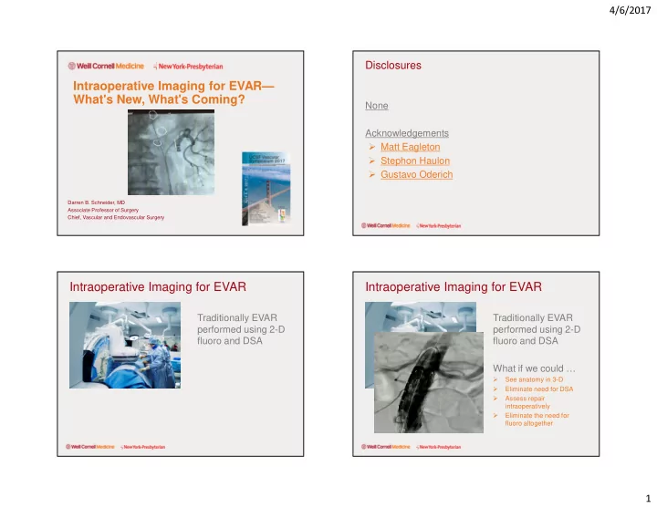
Traditionally EVAR performed using 2-D fluoro and DSA What if we could …
- See anatomy in 3-D
- Eliminate need for DSA
- Assess repair
intraoperatively
- Eliminate the need for
fluoro altogether