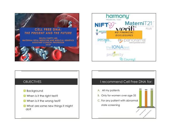
I recommend Cell Free DNA for: OBJECTIVES A. All my patients 34% - - PowerPoint PPT Presentation

i recommend cell free dna for
SMART_READER_LITE
LIVE PREVIEW

I recommend Cell Free DNA for: OBJECTIVES A. All my patients 34% - - PowerPoint PPT Presentation

10/18/2018 N O F I N A N C I A L D I S C LO S U R E S I recommend Cell Free DNA for: OBJECTIVES A. All my patients 34% 34% Background 33% B. Only for women over age 35 When is it the right test? When is it the wrong test? C.


slide-1
SLIDE 1

10/18/2018 1

N O F I N A N C I A L D I S C LO S U R E S

OBJECTIVES

 Background  When is it the right test?  When is it the wrong test?  What are some new things it might do?

I recommend Cell Free DNA for:

  • A. All my patients
  • B. Only for women over age 35
  • C. For any patient with abnormal

state screening

All my patients Only for women over age 35 For any patient with abnormal state sc...

34% 34% 33%

slide-2
SLIDE 2

10/18/2018 2

  • 37-year-old G5P4004 at 12 weeks gestation
  • 3 healthy children
  • 1 son with Wolf-Hirschorn syndrome
  • Parental testing performed
  • Father is a carrier of a balanced translocation

**25% recurrence risk

P a t i e n t W N What testing options should be

  • ffered?
  • A. Cell free DNA
  • B. Invasive testing with CVS
  • C. Cell free DNA followed by CVS
  • nly if abnormal

C e l l f r e e D N A I n v a s i v e t e s t i n g w i t h C V S C e l l f r e e D N A f

  • l

l

  • w

e d b y C V S

  • n

l y i f . . .

13% 26% 61%

C e l l F r e e D N A - w h a t i s i t ?

  • Made up of short

segments of fetal DNA (<200 bp)

  • Origin is primarily

placental in origin

  • Results from apoptosis
  • Circulate in maternal

plasma

cfDNA: Common Clinical Applications

  • Aneuploidy
  • Sex determination
  • Fetal RH status
slide-3
SLIDE 3

10/18/2018 3 Cell Free DNA: Characteristics

cfDNA represents ~10-15% of total DNA in maternal plasma1 Reliably detected after 7 wks gestation2 Higher concentrations late in gestation Short half life (16 min), undetectable by 2 hrs postpartum3

  • JAMA. 2018;320(6):591-592. doi:10.1001/jama.2018.9418

What happens in the lab?

Techniques

  • Massively parallel shotgun sequencing

(MPSS)

  • Targeted sequencing
  • Single nucleotide polymorphisms (SNP)

analysis

slide-4
SLIDE 4

10/18/2018 4

Why does the technique matter?

MPSS Targeted SNP

  • Amplification and

sequencing of all cfDNA

  • Benefit: Broad,

looks at cfDNA from all chromosomes

  • Limitations: Cost,

requires minimum fetal fraction of 4%

  • Sequencing of

regions of interest

  • nly
  • Benefit: less

expensive

  • Comparison of

SNPs of maternal

  • rigin to fetal
  • Benefit: accuracy

is equal across chromosomes

  • May allow

differentiation between triploidy, uniparental disomy, maternal mosaicism

Fetal Fraction

Taglauer, WES, Wilkins-Haug L, Bianchi DW Review: cell-free fetal DNA in maternal circulation as an indicatation of placental health and disease

Fetal Fraction

Prenatal Diagnosis 2012; 32(13) p 1233-41

Why does fetal fraction matter?

The less fetal DNA, the harder to tell normal from abnormal

slide-5
SLIDE 5

10/18/2018 5

Factors that impact fetal fraction

Maternal BMI- increased BMI associated with

decreased FF  Failed to provide a result in 20% of women >250 lb and 50% of women >350 lb.

Gestational age

 Increases after 21 weeks

Aneuploidy

 Increased FF with infants with T21  Decreased FF with infants with T18

Twins Cell free DNA screening: Biologic Challenges

False Positives

  • Unrecognized or “vanishing”twin
  • Placental Mosaicism
  • Low level maternal mosaicism, esp. sex

chromosomes

  • Maternal Malignancy

False negatives

  • Low level of fetal DNA
  • Placental mosaicism
  • Maternal genetic variation (copy

number variants)

Cell free DNA screening: Biologic Challenges

Cell free DNA screening: Biologic Challenges

Failed Results

  • Increased BMI
  • Low level of fetal DNA
  • Fetal Aneuploidy
slide-6
SLIDE 6

10/18/2018 6

CFDNA and aneuploidy: Perf ormance of cf dna

 It is important to counsel patients about the positive predictive value (PPV) of the test  PPV is influenced by prevalence of a disease  An individual PPV can be calculated using maternal age or risk generated from prior screening test  https://www.perinatalquality.org/vendors/ nsgc/nipt/

CFDNA and aneuploidy: Perf ormance of cf dna

PPV calculator

Cell Free DNA for Aneuploidy in Twins

 Unaffected twin may mask an affected twin  Although initial studies are reassuring regarding use of cfDNA for T21 in twins, the overall data are limited  Not currently recommended by ACOG/SMFM  For higher order multiples Maternal age and nuchal translucency measurement for aneuploidy estimation

slide-7
SLIDE 7

10/18/2018 7

Cell Free DNA for fetal sex?

False positive rate is high for monosomy X

 Some studies report rates as high as 91%

Patients may learn about possible sex chromosome aneuploidy even when they are primarily interested in fetal sex In some cases, may be abnormal if the mother has an undiagnosed sex chromosome aneuploidy

Cell free DNA for ultrasound abnormalities?

 Negative cfDNA and isolated ‘soft marker’  Diagnostic testing is not recommended solely for this indication  Structural abnormality on ultrasound  Offer diagnostic testing using microarray  The majority of congenital structural abnormalities are due to microdeletions or single gene disorders that cannot reliably be detected by cell free DNA  Invasive testing is recommended

Cell Free DNA Vs. Serum screening

What if CF DNA is normal followed by abnormal serum screening Raises concern for other chromosomal abnormality In a study by Norton et al. the residual risk

was 2%

The lowest detection rate for cfDNA in women who were <25 years old

Cell free DNA for Rh blood typing?

 Lo and colleagues accurately assess fetal Rh status using cfDNA using PCR  Positive predictive value reported as 93%  Negative predictive value as high as 98%  Moise and colleagues (2016) reported false negative in 1 in 520 cases and inconclusive results in 5-6% of cases

slide-8
SLIDE 8

10/18/2018 8

Cell Free DNA for Microdeletions?

 Chromosomal microdeletions account for many syndromes

 May have developmental delay and medical issues

 There are approximately 40 well described microdeletions  Low baseline prevalence of each microdeletion syndrome

 PPV is signficantly impacted

Microdeletions are rare

Microdeletions are More Common Than Down Syndrome for Women Under 40

Adapted from: Snijders, et al. Ultrasound Obstet Gynecol 1999;13:167–170

Should all wome n be

  • ffere d s cree ning for

microde letions?

slide-9
SLIDE 9

10/18/2018 9

  • Microdeletions can be detected by cfDNA, but…
  • Difficult to validate
  • Much of the data on in vitro samples
  • Clinical testing includes small number of syndromes
  • 22q, 1q36, 5p-, 4p-, 15q11-13 (Prader-

Willi/Angelman)

  • Low positive predictive value

Prevalence of many microdeletion syndromes is unknown

Many such clinical syndromes are caused by different molecular mechanisms

Smaller deletions more difficult to detect

22q syndrome (~1/4000)

85% have 3Mb deletion, 15% smaller “97% detection” refers to only 3Mb deletion

Screening for these microdeletions has not been validated in clinical studies. Routine cell-free DNA screening for microdeletion syndromes should not be performed.

C e l l f r e e D N A t e s t i n g f o r s i n g le g e n e d i s o r d e rs

 Disorders that are a result of DNA changes in a single gene  May affect up to 1% of all pregnancies  Requires identifying fetal DNA that is unique from maternal  These results should not be impacted by placental mosaicism  Current clinically available for limited conditions

 Achondroplasia  Thanatophoric dysplasia  Apert syndrome  Cystic fibrosis

slide-10
SLIDE 10

10/18/2018 10

Case #2: 38 yo G2P1 at 12 weeks What is the next step?

  • A. No further testing needed
  • B. Repeat cell free DNA
  • C. Recommend invasive testing

N

  • f

u r t h e r t e s t i n g n e e d e d R e p e a t c e l l f r e e D N A R e c

  • m

m e n d i n v a s i v e t e s t i n g

11% 82% 7%

T he F u t u r e : N o n i n v a s i v e W h ol e G e no m e S e qu en ci n g

  • Coding and non coding

portions

  • Single nucleotide variants
  • Deletions and Duplications
  • Copy number variants

I s m o re al ways be tt e r ?

  • OBTAIN INFORMATION

ABOUT THE ENTIRE FETAL GENOME

  • RAISES BOTH PRACTICAL

AND ETHICAL ISSUES

  • MAY GIVE

INFORMATION REGARDING RISK FOR ADULT ONSET CONDITIONS THAT ARE NOT RELEVANT

slide-11
SLIDE 11

10/18/2018 11

How to choose a lab

Technique Cost Reporting system Genetic counseling support services

Back to case #1…

 Our patient underwent cell free DNA testing with micro deletions  Her results were normal so she declined any further testing  Level II ultrasound was notable for unilateral cleft lip and palate and abnormal profile  Amniocentesis confirmed deletion of 8.9 MB deletion on 4p consistent with Wolf-Hirshorn

Current guidelines ACOG/SMFM guidelines

Conventional screening is most appropriate first line screen for most patients Ethically any patient may choose cfDNA screening, but should be counseled regarding limitations and benefits Diagnostic testing is required to confirm abnormal results before irreversible decisions Not recommended in twin pregnancies Microdeletion/expanded panels for cell free DNA are not recommended

slide-12
SLIDE 12

10/18/2018 12

Special thanks to Mary Norton, MD & Meg Autry, MD