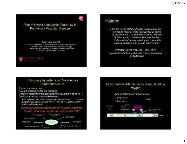
History: Role of Hypoxia Inducible Factor-1 in Pulmonary Vascular - - PowerPoint PPT Presentation

history
SMART_READER_LITE
LIVE PREVIEW

History: Role of Hypoxia Inducible Factor-1 in Pulmonary Vascular - - PowerPoint PPT Presentation

3/11/2017 History: Role of Hypoxia Inducible Factor-1 in Pulmonary Vascular Disease I am convinced that the disease is caused by the increasing mass of inner vascular tissue during its development.to connective tissuecaused


slide-1
SLIDE 1

3/11/2017 1

David N. Cornfield, M.D. Anne T. and Robert M. Bass Professor of Pulmonary Medicine Director-Center for Excellence in Pulmonary Biology Division Director- Pulmonary, Asthma, and Sleep Medicine Stanford University Medical School

Role of Hypoxia Inducible Factor-1α in Pulmonary Vascular Disease

History:

I am convinced that the disease is caused by the increasing mass of inner vascular tissue during its development….to connective tissue…caused by inflammation. However, I contest the term “inflammation” to characterize a process with nothing whatsoever to do with inflammation. Professor Julius Klob, M.D., 1839-1879 regarded as the first to describe primary pulmonary hypertension

Might cell specific expression of hypoxia-inducible factors modulate normal and abnormal PVR?

Pulmonary hypertension: No effective treatment or cure

7 year median survival No cure or reliably effective therapies Despite substantial therapeutic pipeline ($), patient specific Tx Complicates many childhood diseases-

BPD, Primary, CDH, CHD, Immunologic, hypoxia-related pulmonary parenchymal), portal pulmonary HTN, thrombotic, sickle cell, HIV- related, schistosomiasis

HIF-1, 2, 3

Growth, Apoptosis Cell Migration

CXCR4, c-Met

Hormonal regulation

Epo, leptin

Energy Metabolism anaerobic Angiogenesis

VEGF, VEGFR

Vasomotor Tone

ET-1, eNOS,

Matrix, Barrier Function Transport Viral Transcriptional Control

PAS TAD-C TAD-N bHLH

HIF-1α

Hypoxia-inducible factor-1α is regulated by

  • xygen

Two complementary mechanisms:

  • 1. Degradation
  • 2. Inactivation

High O2

O

pVHL

OH OH P402 P564

Ubiquitylation Degradation Ubiquitin Proly-hydroxylases PHD 1,2,3

slide-2
SLIDE 2

3/11/2017 2

PAS TAD-C TAD-N bHLH

HIF-1α

How is HIF-1α regulated by oxygen?

Two complementary mechanisms:

  • 1. Degradation
  • 2. Inactivation

High O2

p300

OH N803

Asparagyl-hydroxylase FIH-1

PAS TAD-C TAD-N bHLH

HIF-1α

Hypoxia-inducible factor-1α is regulated by

  • xygen

Two complementary mechanisms:

  • 1. Degradation
  • 2. Inactivation

High O2

O

pVHL

OH OH P402 P564

Ubiquitylation Degradation Ubiquitin Proly-hydroxylases PHD 1,2,3

Does cell specific expression of HIF-1α modulate lung development?

Altering HIF-1α expression-implications for vascular tone and development

Yu AY, et al. J. Clin. Invest. 1999;103: 691-696

Iyer, et al., Genes&Dev, 1998

HIF+/+ HIF-/-

HIF-1α in pulmonary hypertension- state of the art

Tuder RM, et al. J. Pathol. 2001;195:367-374 Yu AY, et al. Am. J. Physiol. 1998;275:L818-L826 Fijalkowska, I. et al. Am. J. Pathol. 2010. Marsboom G, et al. Circ. Res 2012;110:1484-1497

Control PAH

slide-3
SLIDE 3

3/11/2017 3

SMC specific deletion of HIF-1α increases pulmonary arterial pressure and increases the response to hypoxia

Kim, et al., Circ Res, 2013

SM22-driven deletion of HIF-1α increases myosin light chain phosphorylation in SMC, not muscularization

Kim, et al., Circ Res, 2013

MHC-driven deletion of HIF-1α in SMC decreased pulmonary arterial pressure, but not vascular remodeling

Ball, et al., AJRCCM, 2014

HIF-2α in pulmonary endothelial cells modulates PVR and vascular remodeling

Adult mice 4 month old mice

Tan, et al. JBC, 2013 Tan, et al. JBC, 2013 Cowburn et al. PNAS 2016.

slide-4
SLIDE 4

3/11/2017 4

Demographic Data from control and PH Patients- Pulmonary Hypertension Breakthrough Initiative

IPAH PASMC-P1 Male 40 Hispanic IPAH PASMC-P2 32 Caucasian IPAH PASMC-P3 Male 51 Caucasian IPAH PASMC-P4 Female 27 Caucasian IPAH PASMC-P5 Female 40 Caucasian IPAH PASMC-P6 Female 28 Hispanic IPAH Female Patient ID# Disease Race/Ethnicity Age Gender HT, IH, stroke PASMC-C1 Female 60 Caucasian PASMC-C2 Male Caucasian HT, IH Male 25 Caucasian IH PASMC-C4 Female 49 Caucasian PASMC-C5 Female 57 Caucasian Stroke PASMC-C6 Male 49 Caucasian Control PASMC-C3 IH MI 25

In PA SMC from PH patients, HIF-1α expression is decreased, pMLC is increased

Control IPAH HIF-1α / β-actin

(relative expression) 4 2

**

3.5 1 2.5 1.5 0.5 3

pMLC / MLC

(relative expression) 5 2 6 1

***

3 4

100kD 50kD 37kD 20kD 20kD 1 2 3 4 IPAH 1 2 3 4 Control PASMC HIF-1α β-actin MLC pMLC Barnes, E..A., et al., FASEB, 2017

In PA SMC from PH patients, HIF-1α expression and nuclear translocation are decreased

IPAH Control HIF-1α Barnes, E..A., et al., FASEB, 2017 Nuclei HIF-1α Control IPAH Overlay

+ Normoxic expression of HIF-1α

In pulmonary artery endothelial cells from PAH patients, HIF-1α and HIF-2α expression are increased

Cornfield Lab HIF-1α / β-actin

(relative expression) 3.5 1.5

***

0.5 2.5 2.0 3.0 1.0

50kD 37kD β-actin 100kD HIF-1α 1 2 3 4 IPAH 1 2 3 4 Control PAEC HIF-2α 100kD HIF-2α /β-actin

(relative expression) 1.6 0.8

*

0.2 1.2 1.0 1.4 0.4 0.6

Control IPAH Barnes, E..A., et al., FASEB, 2017

slide-5
SLIDE 5

3/11/2017 5

In PA SMC from PH patients, HIF-1α expression is decreased, pMLC is increased

Barnes, E..A., et al., FASEB, 2017 IPAH Control

pMLC Vinculin pMLC

IPAH Control

In PA SMC from PH patients, prolyl hydroxylase activity is increased

Barnes, E..A., et al., FASEB, 2017

100kD 50kD 37kD HIF-1α N H N H Control IPAH β-actin PHD2 PHD1 PHD3 50kD 50kD 37kD Normoxia Hypoxia HIF-1α / β-actin

(relative expression)

***

Control

14 6 12 2 10 4 8

IPAH

§§§

DMSO DMOG

18 10 16 4 12 6

**

HIF-1α / β-actin

(relative expression)

Control IPAH

2

**

§§§ §§§ 8

Ascorbate

(ng/mL)

*

2 6 16 20 10 12 14 18 4 8

Iron (ng/mL)

0.1 0.7 0.9 0.3 0.5

Fe(II) Fe(III)

***

§§ § IPAH Control 3 2.5

Active MLCK (ng)

2.5 1 0.5

*

2 1.5

Control IPAH

In PA SMC from PH patients, myosin light chain kinase activity and pMLC are increased

20kD 50kD MLCK

  • pMLC

ML7: 37kD β-actin IPAH 20kD MLC Control

  • HIF-1α

100kD 150kD 100kD

***

pMLC / MLC (relative expression)

3.0 2.0 4.0

ML7:

1.0

  • IPAH

Control

  • §

§§

*

Barnes, E..A., et al., FASEB, 2017 12 16 2

Active MLCK (ng)

10 14 6 4 8

**

siNTC siHIF-1α Control IPAH No SMC

100 40 120 80 20

Surface Area (% of no SMC)

60

No SMC IPAH Control

*** *** ***

siNTC siHIF-1α

100 40 120 80 20

Surface Area (% of siNTC)

60

***

Focal Adhesions (per cell) 1h 18h Control IPAH

200 100 250 150 50

*** ***

§§§ §§§ F-actin Vinculin

Control IPAH

In PA SMC from PH patients, contractility is increased which is mimicked by loss of HIF-1α

Barnes, E..A., et al., FASEB, 2017

slide-6
SLIDE 6

3/11/2017 6

20kD 50kD 100kD 37kD 20kD

  • HIF-1α

pMLC β-actin MLC

  • DMOG

Asc

8 2 10 6

HIF-1α / β-actin (relative expression)

***

4

* ***

Non- detectable 3.5 1.5 4.0 2.5 0.5

pMLC / MLC (relative expression)

2.0

*** **

DMOG Asc

  • Surface Area

(% of no SMC)

120 40 20 100 60

**

80

**

Prolyl hydroxylase activity modifies contractility and pMLC Pulmonary vascular cell specific HIF-1-3α expression cells from control patients and PH patients

PASMC +++ HIF-1α HIF-2α HIF-3α Control IPAH + + +

  • PAEC

+ Control IPAH ++ + ++

  • Fibroblasts

N.D. Control IPAH N.D. N.D. N.D. +++ +

HIF likely has cell, temporal and isoform specific effects

Low Tone Pulmonary Artery SMC:

  • HIF-1α present in normoxia
  • Prolyl hydroxylase activity +
  • MLC kinase constrained
  • MLC- P
  • [Ca2+]I limited (KCa subunit)
  • Decreased contractile state

High Tone Pulmonary Artery SMC:

  • HIF-1α decreased in normoxia
  • Prolyl hydroxylase activity ++++
  • MLC kinase elevated
  • MLC P
  • [Ca2+]i increased (KCa subunit)
  • Increased contractile state

PA endothelial cells and SMC hypoxia-inducible factor form and function are distinct

Alveolarization

Secondary Septation Alveolar

Burri PH, Biol Neonate, 2006

Saccular

  • Risk inversely proportional to weight
  • 85% of infants 700g, 30% between 700

and 1000 g.

  • 2/3 of affected infants with only minimal

supplemental O2 or MV

  • 10,000 new BPD cases annually
  • Indeterminate but significant PH

“New BPD” impaired secondary septation and capillary formation, “arrested development” Courtesy of C. Alvira, MD

slide-7
SLIDE 7

3/11/2017 7 Hypoxia-induced HIF-1α protein stabilization is more durable in fetal than adult PA SMC

* p<0.05, vs. normoxia ** p<0.01, vs. normoxia

* * ** ** Fetal PA SMC HIF-1α /tubulin

(relative expression)

* * Adult PA SMC *

Hypothesis: SM-22α HIF-1α expression modulates lung development ?

On C57B/6 Mice, HIF-1α deleted using an SM-22α promoter Tissue Specific, deletion confirmed, no increased neonatal mortality Does expression of HIF-1α in SM22α cells contribute to lung development?

Alveolarization is Compromised in 8 day old and adult SM22α-HIF-1α-/- Mice

SM22α-HIF-1α-/- SM22α-HIF-1α+/+

3d 8d Adult

Mean Linear Intercept (mm) 50

8d Adult

40 20 30 10 60

** **

3d

§§ §§ §§ § Radial Alveolar Count 12 HIF-1α+/+ HIF-1α-/- 4 10 8 2 6 14

8d Adult 3d

** **

§§ §§§ §§ §§§

Length of α-SMC actin positive arteries is decreased in SM-22α HIF-1α-/- mice.

SM22α-HIF-1α+/+ SM22α-HIF-1α-/-

α-SMA

v v v v v a a a a b b b b Pulmonary artery Pulmonary artery

slide-8
SLIDE 8

3/11/2017 8 SM22α expressing cells persist in the lung periphery longer in SM22α-HIF-1α-/- mice

8d 8d

SM22α-HIF-1α+/+ SM22α-HIF-1α-/-

3d 3d SM22α Adult Adult SM22α

Vessel density is decreased in neonatal SM22α-HIF-1α-/- Mice

SM22α-HIF-1α+/+ SM22α-HIF-1α-/-

*

VWF Positive Vessels (per HPF) 8 6 3 5 1 4 2 7 HIF-1α+/+ HIF-1α−/−

VWF

Study Question: How might expression of HIF-1α in SM22α cells influence pulmonary vascular development ? Specifically, during lung development might there be ‘conversation’ between the PAEC and PASMC ?

SMC media starvation media HIF-1α+/+ CM HIF-1α-/- CM

Number of Branch Points (per field) 45 10 40 20 5 15 50 35 30 SMC media HIF-1α+/+ CM HIF-1α-/- CM starvation media

***

25

PAEC tube formation is more well preserved with conditioned media from neonatal HIF-1α+/+ SMC

slide-9
SLIDE 9

3/11/2017 9 PAEC migrate more effectively in response to SM22α HIF-1α+/+ cells

+ HIF-1α+/+ + HIF-1α−/− No SMC Number of Migrated EC (per HPF) 20 40 10 50 30

+ SMC + VEGF

***

+ VEGF + HIF-1α+/+ + HIF-1α−/−

+ SMC + VEGF

*

§§§

Addition of Angiopoietin-2 to conditioned media improves PAEC tube formation and restores migration

HIF-1α-/- CM HIF-1α-/- CM + Ang2 Number of Branch Points (per field) 10 50 20 60 40

***

30

HIF-1α-/- HIF-1α-/- + Ang2

Number of Migrated EC (per HPF) 10 50 20 60 40

***

30

Conclusion:

HIF-1α in the lung:

  • Plays cell-specific roles;
  • Is active even in normoxia;
  • SMC has long-term implications for lung structure and perhaps function;
  • SMC may modulate PAEC migration and angiogenesis and elastin

homeostasis

  • SMC may regulate angiopoietin production to ‘call’ endothelial cells.

Acknowledgements Elizabeth A Barnes, Ph.D.

Lihua Ying, Ph.D. Chihhsin Chen, M.S.

Yumee Kim, PhD.

Saidie Rodriguez, M.D. Ross Metzger, Ph.D. Cristina M Alvira, M.D.