
SLIDE 1
heart MEDIASTINUM The mediastinum is a broad central partition - - PowerPoint PPT Presentation
heart MEDIASTINUM The mediastinum is a broad central partition that separates the two laterally placed pleural cavities . The mediastinum extends: from the sternum to the bodies of the vertebrae ; and from the superior thoracic aperture to
MEDIASTINUM The mediastinum is a broad central partition that separates the two laterally placed pleural cavities . The mediastinum extends: from the sternum to the bodies of the vertebrae; and from the superior thoracic aperture to the diaphragm
The mediastinum is subdivided into several smaller regions BY A transverse plane extending from the sternal angle to the intervertebral disc between vertebrae T 4 and T 5 separates the mediastinum into: 1-SUPERIOR MEDIASTINUM 2-INFERIOR MEDIASTINUM, which is further divided by the pericardial sac into: 1-THE ANTERIOR The area anterior to the pericardial sac and posterior to the body of the sternum . 2-MIDDLE The area in the middle, which includes the pericardial sac and its contents, is the middle mediastinum 3-POSTERIOR MEDIASTINUM The region posterior to the pericardial sac and the diaphragm and anterior to the bodies of the vertebrae is the posterior mediastinum.
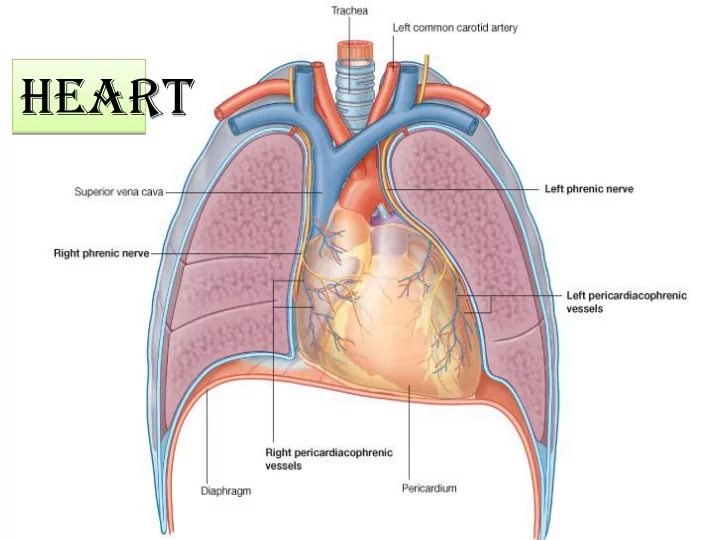
The Pericardium It consists of two components, 1-THE FIBROUS PERICARDIUM : is a tough connective tissue outer layer 2- THE SEROUS PERICARDIUM: is thin layer and consists of two parts: A-The parietal layer lines the inner surface of the fibrous; B- The visceral layer (epicardium)
the heart and forms its outer covering The pericardium is a fibroserous sac surrounding the heart and the roots of the great vessels The narrow space created between the two layers of serous pericardium, containing a small amount of fluid, is the pericardial cavity Pericarditis Pericarditis is an inflammatory condition
The walls of the heart are composed of :
1-EPICARDIUM : visceral serous pericardium 2-MYOCARDIUM: cardiac muscle. 3-ENDOCARDIUM: layer of
endothelium. MYOCARDIUM EPICARDIUM
ENDOCAR DIUM
Heart
shaped
the middlle mediastinum.
Surfaces of the Heart
The heart has three surfaces:
1- Sternocostal (anterior) 2-Diaphragmatic (inferior) 3- A base (posterior) The apex of the heart, formed
by the left ventricle, is directed downward, forward, and to the left It lies at the level of the fifth left intercostal space, 3.5 in. (9 cm) from the midline.
The base of the heart, or the
posterior surface, is formed mainly by the left atrium,
Borders of the Heart The right border is formed by the right atrium; These borders are important to recognize when examining a radiograph of the heart. The lower border is formed mainly by the right ventricle but also by the right atrium; The left border, by the left auricle; and below, by the left ventricle the apex is formed by the left ventricle .
The coronary sulcus circles the heart, separating the atria from the ventricles The posterior interventricular sulcus is on the diaphragmatic surface of the heart The anterior interventricular sulcus is on the anterior surface of the heart and Grooves on its (heart) external surfaces Posterior view Anterior view
Chambers of the Heart The heart is divided by vertical septa into four chambers: 1-the right and left atria 2- the right and left ventricles.
A-Right Atrium
The right atrium consists of a main cavity and a small
Openings into the Right Atrium
1-The superior vena cava :
It returns the blood to the heart from the upper half of the body.
It returns the blood to the heart from the lower half of the body.
3-The coronary sinus
which drains most of the blood from the heart. 4- The right atrioventricular orifice is guarded by the tricuspid valve Fetal Remnants FOSSA OVALIS Located on the atrial septum, which separates the right atrium from the left atrium ; which was the site of the foramen ovale in the fetus
B-Right Ventricle The right ventricle communicates with
1- The right atrium
through the atrioventricular orifice
2-The pulmonary trunk
through the pulmonary orifice As the cavity approaches the pulmonary
point it is referred to as the infundibulum. Its internal structure shows several internal projecting ridges formed of muscle bundles known as trabeculae carneae. An example of trabeculae carneae is the papillary muscles, It is attached by their bases to the ventricular wall while their apices are connected by fibrous chords (the chordae tendineae) to the cusps of the tricuspid valve
C-Left Atrium consists of a main cavity and a left auricle. the fibrous pericardium separates it from the esophagus 1-The four pulmonary veins (two from each lung, 2-The left atrioventricular
Openings into the Left Atrium
4-Left Ventricle
1- The left atrium
through the atrioventricular
2- The aorta through the
aortic orifice
The left ventricle communicates with
The walls of the left ventricle are three times thicker than those of the right ventricle. There are well-developed trabeculae carneae The part of the ventricle below the aortic orifice is called
The aortic vestibule
The aortic valve guards the aortic orifice The pulmonary valve guards the pulmonary orifice and consists of three semilunar cusps. No chordae or papillary muscles are associated with these valves Semilunar valves
The tricuspid valve
fibrous ring of the skeleton of the heart, whereas their free edges and ventricular surfaces are attached to the chordae tendineae.
papillary muscles.
muscles contract and prevent the cusps from being forced into the atrium.
atrioventricular orifice
cusps. Valves are opened and closed by the difference in blood pressure
One of its branches (commonly) is THE POSTERIOR INTERVENTRICULAR (DESCENDING) ARTERY Arterial Supply of the Heart The arterial supply of the heart is provided by : 1-THE RIGHT CORONARY ARTERY 2-LEFT CORONARY ARTERIY which arise from the ascending aorta The right coronary artery:
The left coronary artery is usually larger than the right coronary artery supplies the major part of the heart divides into 1- ANTERIOR INTERVENTRICULAR BRANCH AND 2-A CIRCUMFLEX BRANCH.
Venous Drainage of the Heart Most !blood from the heart wall drains into the right atrium through THE CORONARY SINUS which lies in the posterior part of the atrioventricular groove tributaries of the coronary sinus. GREAT CARDIAC VEIN SMALL CARDIAC VEINS MIDDLE CARDIAC VEINS
THE CONDUCTING SYSTEM OF THE HEART consists of specialized cardiac muscle present in
Conducting System of the Heart
rhythmically at about 70 to 90 beats per minute in the resting adult.
the ventricles allows time for the atria to empty their blood into the ventricles before the ventricles contract.
process originates spontaneously in the conducting system and the impulse travels to different regions of the heart, so the atria contract first and together, to be followed later by the contractions of both ventricles together Read only Read only
The heart is innervated by sympathetic and parasympathetic fibers of the autonomic nervous system via the cardiac plexuses Nerve Supply of the Heart The sympathetic supply arises from the cervical and upper thoracic portions of the sympathetic trunks The parasympathetic supply comes from the vagus nerves Activation of sympathetic nerves results in cardiac acceleration, increased force of contraction of the cardiac muscle, and dilatation of the coronary arteries. Activation of the parasympathetic nerves results in a reduction in the rate and force of contraction of the heart and a constriction of the coronary