
SLIDE 1
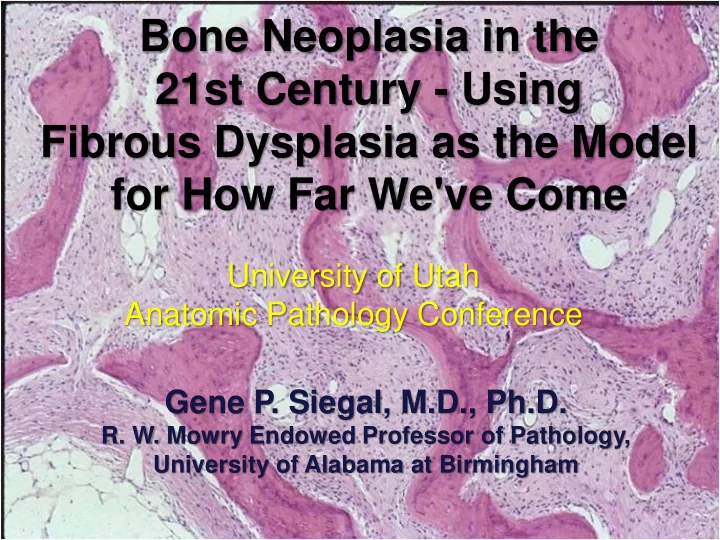
Bone Neoplasia in the 21st Century - Using Fibrous Dysplasia as the Model for How Far We've Come
Gene P. Siegal, M.D., Ph.D.
- R. W. Mowry Endowed Professor of Pathology,
University of Alabama at Birmingham