
SLIDE 1
Polymer Synthesis & Physics Laboratory
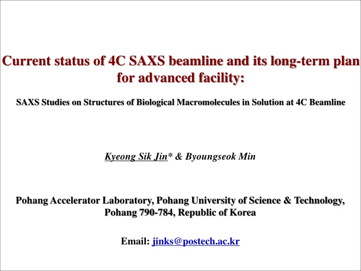
Current status of 4C SAXS beamline and its long-term plan for advanced facility:
SAXS Studies on Structures of Biological Macromolecules in Solution at 4C Beamline
for advanced facility: SAXS Studies on Structures of Biological - - PowerPoint PPT Presentation
Current status of 4C SAXS beamline and its long-term plan for advanced facility: SAXS Studies on Structures of Biological Macromolecules in Solution at 4C Beamline Kyeong Sik Jin* & Byoungseok Min Pohang Accelerator Laboratory, Pohang
Polymer Synthesis & Physics Laboratory
SAXS Studies on Structures of Biological Macromolecules in Solution at 4C Beamline
Small-angle X-ray Scattering (SAXS) is a small-angle scattering (SAS) technique where the elastic scattering of X-rays (wavelength 0.1 ~ 0.2 nm) by a sample which has inhomogeneities in the nm-range, is recorded at very low angle (typically 0.1 ~ 10°). This angular range contains information about the shape and size of macromolecules, characteristic distances of partially ordered materials, pore sizes, and other data. SAXS is capable of delivering structural information of macromolecules between 3 and 25 nm, of repeat distances in partially ordered systems of up to 150 nm. U-SAXS (ultra-small angle X- ray scattering) can resolves even larger dimensions. 4C SAXS beamline is dedicated to conventional transmission small-angle X-ray scattering and diffraction studies for interpreting the structure, structural changes, and relationship between structure and function of molecular/nano structured polymer, self-assembled organic/inorganic nanostructure, composite nanomaterials, biological macromolecules (protein, DNA, and RNA) and their complexes in nearly physiological environments.
Form factor F(q)
Structure factor S(q) local order, relative position
0.0 0.5 1.0 1.5 I(q) S(q)
F(q)
WAXS patterns contain data concerning correlations on an intra-molecular, inter- atomic level SAXS patterns contain data concerning correlations on an inter-molecular level: necessary samples where there is macromolecular or aggregate order As synthesis design/control improves, SAXS becomes more relevant than ever before
Incident beam height = 1400 mm
Beam Size [mm] Beam Size [mm] 1.450 1.650 0.700 0.700 23 um 300 um 0.400 1.030
Undulator
Incidence Angle 2.6 mrad 88.4 mm
Source 0 m DCM 18.0 m FM 21.3 m Defining Slit 11.4 m
Horizontal (33.00 urad) Vertical (29.00 urad) Wall Movable mask Beam stopper
Optical Hutch Experimental Hutch Detector 37 m Front End
(including copper screen/tungsten wire monitors) (including graphite filter)
Beamline Specifications
Beamline 4C SAXS-II
Status Operational Source In-vacuum Undulator 20 (1.4 m short, 20 mm period) Monochromator DCM Si (111) Wavelength 0.06-0.12 nm, currently 0.07 nm Mirror Vertical focusing toroidal, rhodium coated Beam flux 1×10¹² ph/sec Beam size 100(V)×300(H) µm2 Resolution 200 nm ~ 0.3 nm Slits Individually motorized blades of tungsten (W) Sample-to-detector distance 5.0/4.0/3.0/2.0/1.0/0.5/0.2 m Detector Rayonix 2D SX 165 Experimental methods Bulk, solution, liquid crystal, film, powder, sol-gel T-SAXS, T-WAXD User groups 50 user groups (Polymer, bio-,
Research results 40 ~ 50 papers/year Data analysis software ATSAS package, SCATTER, etc. (free download) Staff science SAXS studies for self-assembled nanostructures and biomacromolecules in solution
package for electronic
component and experimental equipment control (Self-development)
and construction
user friendly integrated software package for data measurement and data treatment (Self-development)
stage systems for bulk/powder/film/solution SAXS and WAXD experiments (Self-development)
storage, preparation, treatment, and basic material property analysis
high-throughput application measurements, which is available at 4C beamline
improvement
the performance
4C beamline, including automation of SAXS experiments and data analysis, and time-resolved SAXS setups
study the structure of a wide range of self-assembled nanostructures and biological macromolecules
(MD) simulation that could be combined well with time- resolved SAXS study – Pre-dynamic SAXS studies aimed at the 4th generation XFEL experiment
Future Projects and Goals Previous and Current Status
Bulk, Film (25 ~ 400 ºC)
(-15 ~ 200 ºC)
(25 ~ 400 ºC)
1)Malvern Zetasizer Nano Series DLS, 2)Refrigerated/multipurpose/high speed Centrifuge, 3)Ultarsonic Cleaner, 4)Water Purification System, 5)Cold Storage (4ºC), 6)Ultra-Low Temperature Freezer (-40ºC), 7)Bench Mixer, 8)Hanil Micro-12, 9)Thermo Scientific Heater, 10)METTLER TOLEDO EL204-IC Electronic Balance, 11)Pipettes, 12)S-1700, 13)NanoDrop, 14)GPC-FFF-MALS System
Multi (100 holes) sample stage for powder, bulk, film (25 ºC) Multi (10 lines) sample stage for film (25 ºC) N2 inlet Cooling water inlet Sample cell Heating bar cable Temperature sensor Multi (6 holes) heating sample stage equipped with Eurotherm Controller for powder, bulk, film (25 ~ 400 ºC) Single heating sample stage equipped with Eurotherm Controller for powder, bulk, film (25 ~ 400 ºC)
Quartz-based solution cell Pipette + Tip Quartz capillary heating bar cable Sample cell Julabo connection part Single cooling/heating sample stage for sol-gel (-15 ~ 400 ºC) Single cooling/heating sample stage equipped with Julabo circulation for solution (-15 ~ 200 ºC) Multi (20 holes) cooling/heating sample stage for sol-gel (-15 ~ 200 ºC)
Pump Sample injector GPC column FFF channel UV/Vis MALS (18 channel) FFF RI Fraction collector
[FFF]
1) Injection flow 2) Cross flow 3) Channel flow
Before After
◈ Test Result of BSA Protein Standard (MW = 66.450kDa, C= 2mg/ml, Injection volume = 15 ㎕, Experiment time = 13 min)
Automatic/selective background - transmission corrected 2D data acquisition - 1D radial averaged data acquisition – save, batch processing program buildup
(1) (2) (3) (4) (6) (5)
◈ Bio SAXS P12 beamline at the Petra-Ⅲ storage ring (Leader: Dr. Dmitri Svergun)
Automatic sample changer Purification system Dynamic and movable detector
to purify mixtures of proteins (as requested by user groups)
Synchrotron X-ray Beam Sample 2θ CCD Camera qz qx qy z x y Beamstop )
IMolecules + Buffer solution (q) IBuffer solution (q) IMolecules (q)
▪ Experimental procedures 1 2 3 4 5
0.0 0.1 0.2 0.3 0.4 0.5
q (nm-1)
6 5 4 3 2 1
log I (a.u.)
Solid sphere Hollow sphere Dumbbell Flat disk Long rod D 2 4 6 8 10
r (nm) p(r) (a.u.)
(1) Radial Averaged 1D SAXS Curve (2) Indirect Fourier Transform Method → Pair Distance Distribution Function (PDDF) (3) Ab initio Shape Determination Method → Reconstructed 3D Structural Model
(1) (2) (3)
2D SAXS Pattern
1D SAXS Curve p(r) Function Initial 3D Model Final 3D Model Intermediate 3D Model
Solid sphere Hollow sphere Dumbbell Flat disk Long rod
among solution and crystal and theoretical calculated models
Schematic for SAXS data collection, evaluation, analysis, modeling, and interpretation
Data extrapolation, merging, analysis, modeling and interpretation
Sequence : H2NC6H12-5’-CCC TAA CCC TAA CCC TAA CCC-3’-C6H12(CH2OH)NH2 H+ OH-
Fu Fully lly Closed [contractile ile] ] State : stable le 완전히 닫힌 (수축) ) 상태 Less ss Ope pen [rela laxed] d] State : unstable le 덜 열린 (이완) ) 상태 Less ss Closed [contractile ile] State : unstable le 덜 닫힌 (수축) ) 상태
H+: dissociation OH- : association
Fu Fully lly Open [rela laxed] ] State : stable le 완전히 열린 (이완) ) 상태
Compl plementary ary DNA NA
Ful ullere rene
Representative models used to simulate the structure of i-motif DNA in solution at various pH values.
Fu Fully lly Closed [contractile ile] State 닫힌 (수축) ) 상태 Fu Fully lly Open [rela laxed] ] State 열린 (이완) ) 상태
Strong acidic induced fragmentation
▪ Color lines - Theoretical SAXS curves ▪ Symbols - Experimental SAXS curves ▪ Black solid lines - SAXS curves from the p(r) fit
(a) (b) (c)
pH 6.15
1nm
pH 5.33
1nm
5’ end 3’ end C3 C15
ⅹ
Thymine (T) Adenine (A) Cytosine (C)
pH 8.03
1nm
pH 11.2
1nm
Structural models of the i-motif DNA in solutions of various pH conditions. The reconstructed models were
☞ Improvement o
Less s Closed [contractil ile] State ? ?
(1) Fullerene binding-induced transformation (2) pH-induced conformational switching
Representative models of non-functionalized DNA and fullerene DNA hybrid (FDH) nanomachine and the working switching cycle by protons.
☞ Hy Hydrophobic ic int nteractio ion (소수성 상호작용)
Fu Fully lly Closed [contractile ile] ] State : stable le 완전히 닫힌 (수축) ) 상태 Less ss Open [rela laxed] State : unstable le 덜 열린 (이완) ) 상태 Less ss Closed [contractile ile] State : unstable le 덜 닫힌 (수축) ) 상태
Stabilizing by C60 fullerene : C60 proton-sensitive DNA fullerene DNA hybrid nanomachine
The distance distribution p(r) functions for non-functionalized DNA and FDH in solution at pH ≈ 5 conditions, based on an analysis of the experimental SAXS data.
☞ Improvement of Less ss open [rela laxed] State ?
산성: 분해 H+: dissociation 염기성: 결합 OH- : association
Representative models of i-motif DNA with a carboxyl functionalized C60 and working cycle of the pH-induced conformational switching between the duplex (open state) and the i-motif (closed state) structure in the presence of cDNA.
Fu Fully lly Closed [contractile ile] ] State : stable le 완전히 닫힌 (수축) ) 상태 Fu Fully lly Open [rela laxed] ] State : stable le 완전히 열린 (이완) ) 상태 Comple lementary DNA 상보 DNA
Structural models of duplex conformation of (a, b) i-motif DNA system and (c, d) C60/i-motif DNA hybrid system at different pH values. The reconstructed models were obtained without imposing symmetry restrictions by the program DAMMIN, respectively.
C60 : fullerene Complementary DNA Sample (pH 5.0) Tm (oC) ΔG a (kJ/mol) Power stroke b (%) Closing force c (pN)
i-motif DNA 50.2
80 2.8 C60/i-motif DNA hybrid 65.6
86 8.5
a Van’t Hoff plot b Contraction Strain = Lmax-Lmin/ Lmax c Fclose= Wclose(=ΔG) / ΔL
Sample (pH 8.0) Transition pH Tm (oC) ΔG a (kJ/mol)
Duplex of i-motif DNA system 4.5 51.0
Duplex of C60/i-motif DNA hybrid system 5.5 57.8
cf) Kinesin (2pN), Myosin (3-4 pN)
Step ep 1 Less ess Cl Close
tractile tile] st state Step ep 3 Full lly op
en [rel elaxed] State te Step ep 2 Full lly Cl Clos
tile] st state
Structural models of porcine pepsin at different pH values. The reconstructed models were obtained without imposing symmetry restrictions by the program GASBOR, respectively.
Jin, K. S.; Yoon, J.; Heo, K.; Jin, S.; Kim, J.; Kim, S; Ree M, J. Phys. Chem. B 2008, 112, 15821-15827.
(i)
Collaboration : Dr. Eunice EunKyeong Kim (KIST)
Jin, K. S.; Park, J. K.; Yoon, J.; Rho, Y.; kim, J. H.; Kim, E.; Ree M, J. Phys. Chem. B 2008, 112, 9603-9612.
Collaboration : Prof. Kyeong Kyu Kim (Sungkyunkwan Univ.)
(1) Kim, D. Y.; Jin, K. S.; Kwon, E.; Ree, M.; Kim, K. K. Proc. Natl. Acad. Sci. U.S.A. 2007, 104, 8779-8784. (2) Jin, K. S.; Kim, D. Y.; Rho, Y.; Le, V. L.; Kwon, E.; Kim, K. K.; Ree. M. J. Synchrotron Rad. 2008, 15, 219-222.
(a) Crystal structure of Escherichia coli RseB at a resolution of 0.24 nm The solution models of RseB (b) and RseA121–216/RseB complex (c) restored from the SAXS data at a resolution of 1.25 nm. The ribbon diagram of the RseB is overlapped onto the solution model of RseB for the comparison of overall shape and dimension
Science of advanced Materials 6, 2325 (2014)
Yaoyao Fu, Youngran Kim, Kyeong Sik Jin, Hyun Sook Kim, Jong Hyun Kim, DongMing Wang, Minyoung Park, Chang Hwa Jo, Nam Hoon Kwon, Doyeun Kim, Myung Hee Kim, Young Ho Jeon, Kwang Yeon Hwang, Sunghoon Kim, Yunje Cho Proceedings of the National Academy of Sciences of the United States of America 2014, 111, 15084-15089.
[ Hexameric structure of the RQA subcomplex ] “Sturcture of the ArgRS-GlnRS-AIMP1 complex and its implications for mammalian translation”
“Molecular basis for SMC rod formation and its dissolution upon DNA binding”
Young-Min Soh, Frank Bürmann, Ho-Chul Shin, Takashi Oda, Kyeong Sik Jin, Christopher P. Toseland, Cheolhee Kim, Han-Sol Lee, Su Jin Kim, Min-Seok Kong, Marie-Laure Durand-Diebold, Yeon-Gil Kim, Ho Min Kim, Nam-Ki Lee, Mamoru Sato, Byung-Ha Oh*, Molecular Cell 2015, 57, 290-303.
Collaboration : Prof. Kyeung-Jin Kim (Kyungpook National Univ.) Jae-Woo Ahn†, Kyeong Sik Jin†, Hyeoncheol Francis Son, Jeong Ho Chang, and Kyung-Jin Kim* Scientific Reports 2015, 5, 14251
Collaboration : Prof. Eun Chul Cho (Amorepacific & Hanyang Univ.) Do-Hoon Kim, Sora Lim, Jongwon Shim, Ji Eun Song, Jong Soo Chang, Kyeong Sik Jin*and Eun Chul Cho* ACS Applied Materials & Interfaces 2015, 7, 20438-20446
Sample NCPMs CP CPMs Mo Model sphere sphere Typ ype homogeneous core e and and shell ll homogeneous core e and and shell ll
aRc (nm)
3. 3.22 220
b(4.
4.317) 1. 1.46 460
b(1.
1.914)
cσR
0. 0.32 326
0.32 322
2. 2.88 888
e(3.
3.944) 1. 1.22 228
e(1.
1.732)
fRm (nm)
11 11.9 .90
g(15
15.69) 13 13.7 .70
g(17
17.96)
hRm, max (nm)
10 10.6 .61
i(14
14.48) 12 12.2 .21
i(16
16.40)
jRho
0. 0.10 106
0.01 019
Hyun-Chul Kim*, Kyeong Sik Jin*, Se Guen Lee, Eunjoo Kim, Sung Jun Lee, Sang Won Jeong, Seung Woo Lee, and Kwang-Woo Kim. Journal of Nanoscience and Nanotechnology 2016, 16, 6432-6439.