Foot and Ankle Exam Founder, SportZPeak Inc. Sanofi, Investigator - - PowerPoint PPT Presentation

foot and ankle exam
SMART_READER_LITE
LIVE PREVIEW

Foot and Ankle Exam Founder, SportZPeak Inc. Sanofi, Investigator - - PowerPoint PPT Presentation

12/10/2016 Disclosure Founder, RunSafe, RaceSafe Foot and Ankle Exam Founder, SportZPeak Inc. Sanofi, Investigator initiated grant Anthony Luke MD, MPH, CAQ (Sport Med) ABC s of MSK Care UCSF Sports Medicine December 10,


slide-1
SLIDE 1

12/10/2016 1

Foot and Ankle Exam

Anthony Luke MD, MPH, CAQ (Sport Med) UCSF Sports Medicine ABC’s of MSK Care December 10, 2016

Disclosure

  • Founder, RunSafe™, RaceSafe™
  • Founder, SportZPeak Inc.
  • Sanofi, Investigator initiated grant

Outline

  • Anatomy of the Ankle
  • Anatomy of the Foot
  • Common ankle problems
  • Common foot problems
  • Examination of the foot and ankle
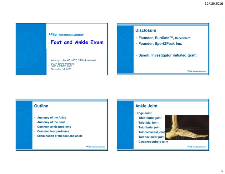
Ankle Joint

Hinge Joint

  • Tibiofibular joint
  • Talotibial joint
  • Talofibular joint
  • Talocalcaneal joint
  • Talonavicular joint
  • Calcaneocuboid joint
slide-2
SLIDE 2

12/10/2016 2

Ankle Movements

Mortise

  • Dorsiflexion, plantarflexion

Subtalar joint

  • Inversion eversion
  • Moves like an angled hinge

Ankle Ligaments

Lateral Collateral Complex

  • Anterior talofibular lig.
  • Calcaneofibular lig.
  • Posterior talofibular lig.

Medial Ligament Complex

  • Deltoid ligament
  • Superficial and deep ligaments

Ankle Ligaments

Sinus tarsi / Subtalar joint

  • Interosseous talocalcaneal ligament

Tibiofibular joint

  • Anterior and posterior tibiofibular ligaments,

intraosseous membrane

Muscles

Posteromedial

“ “ “Tom, Dick And Not Harry” ” ” ”

  • Posterior Tibialis, Flexor

Digitorum, Tibial Artery,Tibial Nerve, Flexor Hallucis Longus

slide-3
SLIDE 3

12/10/2016 3

Muscles

Lateral

  • Peroneus Longus, Brevis, Tertius

Superior

  • Anterior tibialis, extensors

Posterior

  • Achilles Tendon

Anatomy of The Foot

  • Hindfoot
  • Chopart’

’ ’ ’s Joint

  • Midfoot
  • Lisfranc’

’ ’ ’s Joint

  • Forefoot

Acute Ankle Injuries

Mechanism of injury

  • Inversion (Supination)
  • Eversion (Pronation)
  • Axial load
  • Muscle rupture

Position of foot

  • Dorsiflexed or plantarflexed

Ankle Sprains

  • Inversion, plantarflexion is most common

injury

slide-4
SLIDE 4

12/10/2016 4

Associated Injuries

  • Subtalar joint sprain
  • Sinus tarsi syndrome
  • Osteochondral talus

injury

  • Lateral talar process

fracture

  • Posterior impingement

(os trigonum)

  • Posterior tibial tendon

strain

  • Peroneal tendon

subluxation

  • Fracture at the base
  • f the fifth metatarsal
  • Jones fracture
  • Syndesmotic sprain
  • Salter fracture (fibula)
  • Ankle fractures

Ottawa Ankle Rules

  • Inability to weight bear immediately and in the

emergency / office (4 steps)

  • Bone tenderness at the posterior edge of the

medial or lateral malleolus (Obtain Ankle Series)

  • Bone tenderness over the navicular or base of

the fifth metatarsal (Obtain Foot Series)

  • Sens 97%, Spec 31-63%, NPV 99%, PPV <20%

(Am J Emerg Med 1998; 16: 564-67)

Grading Ankle Sprains

  • Grade 1 = mild stretch with no instability
  • Grade 2 = mild laxity with a good end point

(ATFL usually torn but CFL and PTFL intact)

  • Grade 3 = laxity poor end point (ATFL and CFL

usually torn)

Treatment of Ankle Sprains

Acute

  • Ice, Compression, Elevation
  • Rest or modified activities
  • Crutches PRN
  • Bracing (Grade 2 and 3)
  • Physical Therapy
  • Early Motion is essential
slide-5
SLIDE 5

12/10/2016 5

Prognosis

  • Grade 1 – 2 to 4 weeks
  • Grade 2 – 4 to 6 weeks
  • Grade 3 – 6 to 12 weeks
  • More than 12 weeks – consider associated

injuries causing chronic ankle pain

Foot issues

  • Morton’s Neuroma
  • Bunions
  • Hallux Rigidus
  • 2nd metatarsalgia
  • Plantar fasciitis

MRI Plantar Fasciitis Plantar Fasciitis Injection

Patient Supine Landmarks:

  • Medial tubercle of the

calcaneus (insertion of plantar fascia into the calcaneus) is usually the point of maximal tenderness

  • Go 2 cm distal to the most

tender point then aim needle toward tender point

slide-6
SLIDE 6

12/10/2016 6

Plantar Fascia Injection #2

  • Alternative

Technique: Injection approach through the medial heel border posterior to the point

  • f heel tenderness
  • Avoid inferior

approach

Plantar Fascia Injection

  • Post injection:
  • Check pre and post injection pain levels with walking and

palpation (injection can be diagnostic)

  • Lidocaine effect should be immediate and the steroid effect

will occur in 24 hours

  • Continue other treatments (i.e. stretching, NSAIDS,

footwear, +/- nightsplint, etc.)

  • Decrease physical activity for 5-10 days after injection

Plantar Fascia Injection

  • Complications
  • Bleeding, Infection
  • Plantar fascia

rupture

  • Fat pad atrophy

Posterior Tibilialis (PT) Dysfunction

  • Main dynamic stabilizer of

the hindfoot to prevent Heel EVERSION

  • Hyperpronation
  • Tendon dislocation or

rupture (partial vs Complete tears) can cause flatfoot deformity

slide-7
SLIDE 7

12/10/2016 7

PT Dysfunction

Presentation

  • Usually women > 40 y.o.
  • Weakness with inversion

“ “ “Too many toes sign” ” ” ”

  • Unable to plantarflex – Single limb heel

rise test

PT Dysfunction Subtalar neutral Orthotics

  • Material

(polypropylene, cork, rigid plastic)

  • Length (3/4 or full)
  • Lift
  • Posting (forefoot,

midfoot, rearfoot)

Courtesy of Alicia Knee, DPM, UCSF

slide-8
SLIDE 8

12/10/2016 8

Screening Orthotics

Courtesy of Alicia Knee, DPM, UCSF