exercise and bone mass
play

Exercise and Bone Mass Systematic Reviews of RCTs & - PDF document

Clinical Decision Making for Standing June 2003 Programs for Children with Disablities Pyramid of Evidence Exercise and Bone Mass Systematic Reviews of RCTs & Meta-Analysis Whats the Evidence? Randomized Controlled Clinical Trials (RCT)


  1. Clinical Decision Making for Standing June 2003 Programs for Children with Disablities Pyramid of Evidence Exercise and Bone Mass Systematic Reviews of RCTs & Meta-Analysis Whats the Evidence? Randomized Controlled Clinical Trials (RCT) Is there a mechanism? Cohort Studies Case-Controlled Studies Clifford J Rosen MD Maine Medical Center
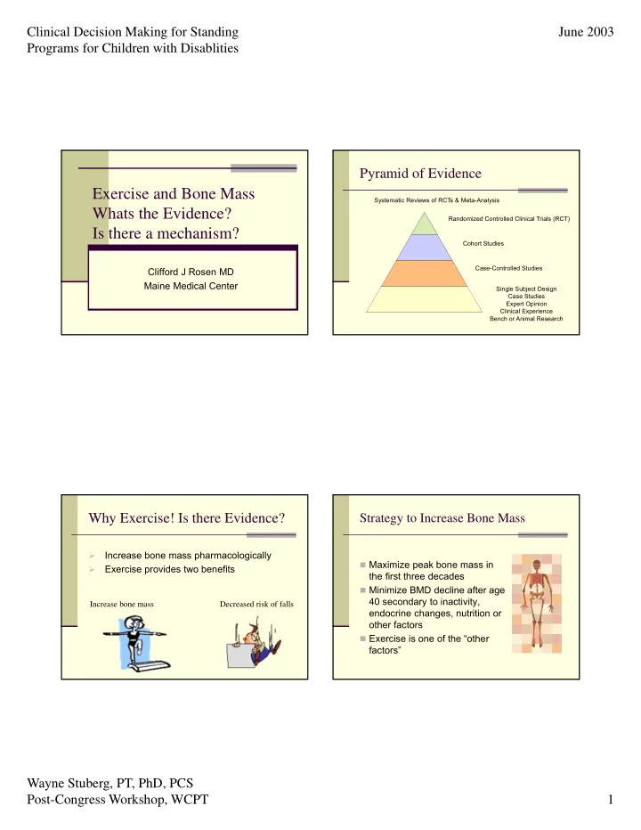
 Single Subject Design Case Studies Expert Opinion Clinical Experience Bench or Animal Research Why Exercise! Is there Evidence? Strategy to Increase Bone Mass Increase bone mass pharmacologically   Maximize peak bone mass in  Exercise provides two benefits the first three decades  Minimize BMD decline after age 40 secondary to inactivity, Increase bone mass Decreased risk of falls endocrine changes, nutrition or other factors  Exercise is one of the “other factors” Wayne Stuberg, PT, PhD, PCS Post-Congress Workshop, WCPT 1

  2. Clinical Decision Making for Standing June 2003 Programs for Children with Disablities Bone Health and Exercise: Too Little and Too Much- Are They Exercise and Bone Mass Controversy Related?  Overall role of exercise in prevention and Optimal Bone Health treatment of osteoporosis is unclear  Pros  Absence of mechanical loading, gravity has been shown to cause a decrease  Active individuals have greater bone mass than sedentary individuals as shown in cross- sectional studies Too Little – Too Much –  Bone mass increases with activity in sedentary osteomalacia Stress fractures individuals in longitudinal studies & osteopenia or amenorrhea Zero gravity Exercise Is Not the Path to Strong Bones By GINA KOLATA APRIL 1, 2016 Photo Exercise and Bone Mass Controversy Moderate exercise has many benefits, but it does not build strong bones. CreditTara Moore/Getty Images Misconception: All you have to do is walk or do even the most modest strength training exercises to build strong bones.  Controversy  Bone mass development and maintenance is multifactorial with nutrition and hormonal influences that can have a greater impact  Exercise prescription is unclear  No dose-response relationship has been confirmed in the developing skeleton nor Exercise Is Not the Path to Strong Bones for either the middle aged nor elderly Wayne Stuberg, PT, PhD, PCS Post-Congress Workshop, WCPT 2

  3. Clinical Decision Making for Standing June 2003 Programs for Children with Disablities NASA is currently working on an Exercise Countermeasures Project to help promote the The astronauts do treadmill health and safety of astronauts and reduce the exercises that include walking, effects of zero gravity. running, deep knee bends, and They are studying and developing exercises that resistive exercises. These exercises are designed to astronauts can do while in space. They are stimulate bone mass, working to come up with equipment and workouts cardiovascular fitness, muscle that are both time and space efficient. Some of endurance, and the the products of this program are depicted in the neurophysiologic pathways and next few slides. reflexes required for walking on Earth or other planetary surfaces. What is he doing? * The astronaut depicted to the right is doing “resistive training.” He is strapped into the device and weight is imparted on his body to help simulate the weight he would normally experience while on earth. This particular machine can impart up to 300 lbs of pressure. The astronaut on the left is doing cycle ergometry. This exercise helps to improve endurance and strengthen her bones when resistance is Image from Nasa webpage http://www.nasa.gov/mission_pages/station/science/eZLS_treadmill_010306.html adequately applied . Wayne Stuberg, PT, PhD, PCS Post-Congress Workshop, WCPT 3

  4. Clinical Decision Making for Standing June 2003 Programs for Children with Disablities Does Exercise Affect Bone Mass in Developing Optimal Bone Mass Infants?  If exercise is beneficial then a number of Yes, 5 randomized clinical trials provide questions arise: evidence to support enhanced bone development in children using exercise as an  When and how do I begin developing optimal intervention: bone mass? •Moyer-Mileur et al, 1995  Is exercise in infancy or prepuberty advised? •Moyer-Mileur et al, 2000  Is exercise needed to be continued as part of •Nemet et al, 2001 a person’s lifestyle into adulthood? •Litmanovitz et al, 2003  How much is enough? •Aly et al, 2004 BAT is Related to Muscle Mass in Infants Boxplots of MRI measures of BAT in the supraclavicular area and paraspinous musculature in 30 infants. While the proportion of BAT in the supraclavicular area decreased, measures of paraspinous musculature increased from birth to six months of age; both P ’s < 0.001 (Ref). Below is an example of an MRI fat fraction (FF) image of a female infant at 2 days and 6 months of age showing a marked decrease of BAT in the supraclavicular area (arrows), as indicated by an increase in FF. Wayne Stuberg, PT, PhD, PCS Post-Congress Workshop, WCPT 4

  5. Clinical Decision Making for Standing June 2003 Programs for Children with Disablities BAT May Influence Skeletal Acquisition in Infancy Exercise & Bone Mass in Prematurity  Random assignment of 32 preterm infants  Birth weight of 800-1600 g & 26-32 wks gestation  Stratified assignment using birth weight & gestational age then randomly assigned to exercise or control  4 week exercise program  5 repetitions of passive range of motion (ROM) with mild manual compressions to the wrist, elbow, shoulder, ankle, knee & hip  5-10 minutes in duration, 5 days per week  Control program with holding & stroking Moyer-Mileur et al, 2000 Exercise & Bone Mass in Prematurity Exercise & Bone Mass in Prematurity  Random assignment of 24 preterm infants  Significant difference between groups for  ≈ 1000 gr, 28 wks gestation, corrected age of serum type I collagen C-terminal propeptide 33 weeks at enrollment (correlates with collagen turnover and bone formation in premature infants)  4 week exercise program  Significant difference between groups for  5 repetitions of passive ROM with mild manual urine pyridinoline (marker for bone resorption) compressions to the wrist, elbow, shoulder,  Significant increase in DEXA measures of: ankle, knee & hip  forearm length & bone area,  5-10 minutes in duration, 5 days per week  BMC & BMD  Control program with holding & stroking Nemet, et al, 2001 Wayne Stuberg, PT, PhD, PCS Post-Congress Workshop, WCPT 5

  6. Clinical Decision Making for Standing June 2003 Programs for Children with Disablities Exercise & Bone Mass in Children and Adolescents Exercise & Bone Mass in Prematurity E f f e c t o f P a s s iv e R O M  Research evidence supports higher bone mass in children who participate in 4 0 activities with high impact forces than 3 0 sedentary controls: 2 0 1 0  Jumping ( MacKelvie et al, 2002 & 2003, Fuchs et al, % C h a n g e C o n t r o l 0 2001, Pettit et al, 2002, Johannsen et al, 2003 ) E x e r c i s e - 1 0  Tennis ( Haapasalo et al, 1996, Bass et al, 2002) - 2 0  Gymnastics (Zanker et al, 2003) - 3 0  Weight Lifting ( Nichols et al, 2001 ) B S A P P I C P I C T P  General exercise with impact loading and Significant increase in bone-specific alkaline phosphatase (BSAP), increase in C- strengthening ( Morris et al, 1997, McKay et al, 2000, terminal procollagen type 1 peptide & significant decrease in C-terminal type- Specker & Binkley, 2003 ) 1collagen telopeptide (ICTP) Jumping & Bone Mass Jumping & Bone Mass  School-based Jumping in Girls (Petit et al, 2002)  Results  14 schools in Canada with 383 girls  No significant bone structure change in  Grades 4-6 (ages 9-12 years) prepubertal girls (Tanner stage 1)  7 month program of jumping, 10 min/day, 3 times per  Significant bone structure change in early week vs. regular activities pre-pubertal (Tanner stage 2-3)  Rated maturity with Tanner Scale  Increase bone cross-sectional area & section  Measured calcium intake & general physical activities modulus due to less endosteal resorption & increased cortical thickness  DEXA scans of femoral neck, trochanteric & proximal shaft with calculation of:  No significant difference in periosteal width  Bone cross-sectional area  Cross sectional moment of inertia  Section modulus (indicator of bone strength) Wayne Stuberg, PT, PhD, PCS Post-Congress Workshop, WCPT 6

Download Presentation
Download Policy: The content available on the website is offered to you 'AS IS' for your personal information and use only. It cannot be commercialized, licensed, or distributed on other websites without prior consent from the author. To download a presentation, simply click this link. If you encounter any difficulties during the download process, it's possible that the publisher has removed the file from their server.

Recommend


More recommend