Evaluate the vitreoretinal interface routinely. - - PDF document

evaluate the vitreoretinal interface routinely
SMART_READER_LITE
LIVE PREVIEW

Evaluate the vitreoretinal interface routinely. - - PDF document

Financial Disclosure: JP CNV Variations and Honoraria Alcon Notal Vision Masqueraders Reichert Scientific Advisory Boards Zeavision Carl Zeiss Meditec Carlo J. Pelino, OD, FAAO Proprietary Interests


slide-1
SLIDE 1

1

CNV Variations and Masqueraders

Carlo J. Pelino, OD, FAAO Cpelino@salus.edu Joseph J. Pizzimenti, OD, FAAO pizzimen@nova.edu

Financial Disclosure: JP

  • Honoraria

– Alcon – Notal Vision – Reichert

  • Scientific Advisory Boards

– Zeavision – Carl Zeiss Meditec

  • Proprietary Interests

– None

  • CEO/Founder

– Optometryboardcertified.com

  • CE Companies

– CEinItaly.com – EyeSkiUtah.com

Financial Disclosure: CP

  • CE Companies

– CEinItaly.com

  • Dr. Pelino has

no proprietary interests or other financial relationships to disclose.

Course Goal

  • To provide useful clinical information about

variations and masqueraders of CNV.

– Classification, diagnosis, treatment/management

Evaluate the vitreoretinal interface routinely.

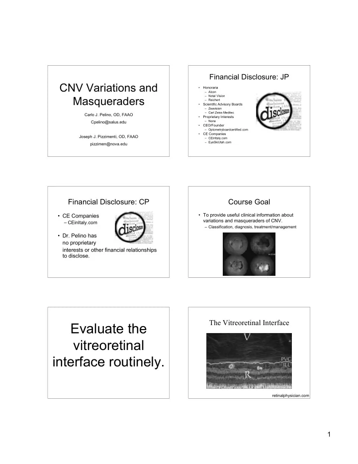
The Vitreoretinal Interface

retinalphysician.com

slide-2
SLIDE 2

2

Persistent Vitreomacular Adhesions (VMA)

Anomalous PVD Anomalous PVD

  • May hasten the Wet

AMD process. ---->

The Retina

  • RPE
  • Neurosensory
  • 6 million Cones
  • Detailed vision
  • Color vision
  • 120 million Rods
  • Peripheral retinal

receptors

  • Great sensitivity to

light

OS IS/OS ELM RPE IS NFL: Nerve Fiber Layer OPL: Outer Plexiform Layer IS/OS: Junction of inner and outer ILM: Inner Limiting Membrane ONL: Outer Nuclear Layer photoreceptor segments GCL: Ganglion Cell Layer ELM: External limiting membrane OS: Photoreceptor Outer Segment IPL: Inner Plexiform Layer IS: Photoreceptor Inner Segment RPE: Retinal Pigment Epithelium INL: Inner Nuclear Layer ILM GCL NFL Choroid IPL INL OPL ONL

SD-OCT Healthy Macula Jetrea (ocriplasmin)

slide-3
SLIDE 3

3

Retina

  • The Pigment Epithelium

– Monolayer – Cuboidal cells – Function of RPE – Tight junctions form outer blood-retina barrier

RPE Retinal Pigment Epithelium

  • 120 million cells in

monolayer

  • Functions of RPE

– Phagocytosis of renewable discs of PRs – O-2 diffusion to PRs – Provision of nutrients to PRs

Early AMD: Accumulation of Lipofuscin and Vitamin A Metabolites

Reduced degradation of cellular debris leads to the accumulation of lipofuscin, toxic vitamin A metabolites

Drusen Retina

  • The Neurosensory Retina

– The Photoreceptors

  • Structure and function of cones and rods

– Inner and outer segment junction

  • Importance of structural integrity to visual function

– Outer limiting membrane – Outer nuclei – Synaptic layer (plexiform)

slide-4
SLIDE 4

4

Photoreceptors Neuro-sensory Retina

  • Inner nuclei
  • Synaptic layer

(plexiform)

  • Ganglion cells
  • Nerve fiber layer
  • Internal limiting

membrane

Retinal Vasculature

  • 2 main sources of blood

supply:

  • Choroidal BV

– Supplies outer retinal layers, including PRs

  • CRA

– 4 branches nourish inner retina – Run radially toward fovea

Retinal Capillaries

  • Pericytes surround

each endothelial cell – provide support

  • Tight junctions between

endothelial cells

  • Pericytes + tight

junctions form inner blood-retinal barrier. Pericytes marked by ng2 staining (blue) and endothelial cells are marked by PECAM (red).

Retina

  • Photransduction

– conversion of light into an electrical impulse

  • The retina is damaged by it’s own
  • peration.
  • Autoregulation of blood flow
slide-5
SLIDE 5

5

Functional Anatomy: The Fovea The Choroid

  • Vascular layers
  • Melanocytes
  • Bruch’s membrane
  • Sympathetic regulation of blood flow
  • Function of choriocapillaris

– Supply of nutrients – Absorption of light

Diagnostic Dilemma

Choroidal Neovascularization

Angiogenesis

Environmental factors1 (hypoxia,2 pH) Growth factors, hormones1 (EGF, bFGF, PDGF, IGF-1, IL-1!, IL- 6, estrogen) VEGF-A binding and activation of VEGF receptor3 Endothelial cell activation3

VEGF-A = vascular endothelial growth factor A; EGF = epidermal growth factor; bFGF = basic fibroblast growth factor; PDGF = platelet-derived growth factor; lGF = insulin-like growth factor; IL= interleukin.

  • 1. Dvorak HF. J Clin Oncol. 2002;20:4368. 2. Aiello LP, et al. Arch Ophthalmol. 1995;113:1538.
  • 3. Ferrara N, et al. Nat Med. 2003;9:669. 4. Griffioen AW and Molema G. Pharmacol Rev. 2000;52:237.

CNV ---> FV Scar

slide-6
SLIDE 6

6

CNV has several variations, causes, and masqueraders. Choroidal Neovascularization

  • Subjective symptoms
  • Objective data
  • Diagnostic Workup
  • Making the diagnosis

Common Causes of CNV

  • Exudative AMD
  • Ocular Histoplasmosis
  • High Myopia
  • Angioid Streaks

Fundus Autofluorescence Fundus Autofluorescence

Fundus Autofluorescence Wet AMD

slide-7
SLIDE 7

7

Fluorescein Angiography

Indocyanine Green Angiography (ICGA)

  • Uses digital imaging systems
  • Dye properties
  • “Sees” through blood
  • Delineates choroidal circulation better than

fluorescein angiography

  • Boundaries of occult membranes imaged

Occult CNV Classic CNV Type I CNVM

Retina RPE Bruch’ s CC

CNV beneath RPE (AMD) *

Fovea Retina RPE Bruch’s

Type II CNVM

CNV in subsensory space (POHS)

slide-8
SLIDE 8

8

ANGIOID STREAKS

  • Note Angioid Streaks radiating from the optic discs and macular

laser scarring

Differential Dx. of Angioid Streaks: PEPSI Causes of CNV

  • High Myopia in a 52

y/o WM

  • CNV w/heme
slide-9
SLIDE 9

9

48 y/o WM

  • 12.00D

Concave fundus, CNV, schisis Causes of CNV

  • OHS

Case

! 81 Year old Female with a history of

arthritis.

! 7 year history of injections with

Avastin or Lucentis

! PMH: AMD OU, Cataracts OU ! OcHx: Injections for AMD.

Ophthalmic Exam

! VA:

  • OD: 20/400

OS: 20/80

! IOP

  • OD: 11

OS: 12

! SLE:

  • OD: NS +1

OS: NS + 1

  • DFE:
  • PED OD and Geo Atrophy OS

OCT After Switching to Eyelea

slide-10
SLIDE 10

10

Comparison

!"#$%&'"()"*+%',-"%-. /01$-,' 234533 2"6-7$1,(-'"()"89$:$% 234;3

Another PED example Variations and Masqueraders

  • f CNV

! Polypoidal Choroidal Vasculopathy (PCV) ! Retinal Angiomatous Proliferation (RAP) ! Masqueraders of CNV

  • Choroidal Neoplastic Disease
  • Primary Tumors of the Choroid

! Nevus vs. melanoma

  • Metastatic Tumors to the Choroid

! Common primary sites ! Breast ! Lung

  • Central Serous Chorioretinopathy (CSC)

CNV Variants

R C

ICG Angiography AMD vs PCV

AMD -SUBRETINAAL HEMORRHAGE SUSPICIOUS POLYPOIDAL

slide-11
SLIDE 11

11 Retinal Angiomatous Proliferation

! Sub retinal neovascularization ensues.

Retinal Angiomatous Proliferation

! First described by Yannuzzi in 1991,

RAP is a retino-choridal anastamosis.

! Intraretinal capillary proliferation,

which extends throughout the sensory retina and then into the sub retinal space.

Retinal Angiomatous Proliferation

! 10-20 % of neovascular AMD

patients start with RAP.

! The age group is thought to be

slightly older.

! ICGA aids in confirming diagnosis,

identifying “hot spots” of ICG dye pools in the sub retinal space.

Retinal Angiomatous Proliferation “Hot Spot” Retinal Angiomatous Proliferation

82 y/o WM w/drusen

slide-12
SLIDE 12

12

RAP Stage I: intraretinal neovascularization. RAP Stage II: subretinal NV w/retinal-retinal anastomosis. RAP Stage III: subretinal NV w/vascularized RPED and retina-choroid anastomosis.

slide-13
SLIDE 13

13 RAP: Current Treatment Options

! Thermal Laser ! Photodynamic Therapy ! Anti VEGF Therapy

CNV Masquerador: Neoplastic Disease CNV or Mass? CNV Masquerador:

Central Serous Chorioretinopathy

Mystery Macula

! Subjective

  • 35 y/o WM
  • sudden, unilateral blur OD
  • no pain or trauma
  • “Type A”

! Objective

  • VA

! OD 20/60 ! OS 20/20

  • Hyperopic shift
slide-14
SLIDE 14

14

Describe That Fundus!

! DFE shows large, serous elevation ! Focal detachment of sensory retina

What other tests would you like to perform?

OCT

slide-15
SLIDE 15

15

(Idiopathic) Central Serous Chorioretinopathy

(ICSC)

Patient Outcome

  • VA recovered to 20/25

at week 12

  • Reduction of fluid,

20/40 VA at week 5

Central Serous Chorioretinopathy

! 36 y/o WM ! CC: Sudden

central blur OS

! VA OD 20/20 ! VA OS 20/200

ICSC

! Objective

  • Breakdown of outer blood-retina barrier
  • FA shows classic “smoke-stack”

! Pooling beneath RPE detachment ! Dye ascends vertically, then laterally in SRS

! Differential Diagnosis

  • Tumor
  • RPE detachment/CNVM
  • Steroid-induced CSC
slide-16
SLIDE 16

16

ICSC Plan

! Observation

  • 60% regain 20/20 w/no intervention
  • monitor q4wks for 6 mon

! Focal Laser

  • Unresolved after 4-6 mon
  • Recurrent
  • Focal, direct treatment
  • Leak must be outside FAZ (500 um)

Treatments for CSC

! Thermal laser ! Photodynamic Therapy

  • Visudyne (Verteporfin)
  • A light-activated drug

Photodynamic Therapy for CSC

! Serous

detachment before PDT. Resolution of detachment with residual RPE mottling after PDT.

What’s new in CSC Treatment?

! Intravitreal

bevacizumab (Avastin) has shown some benefit in small case series.

Low-fluence PDT

ICGA-guided, lower flow, lighter dosage resulted in less hypoperfusion of the choriocapillaris

slide-17
SLIDE 17

17

Current and Future Treatment of CNV

Laser

! Now reserved for extrafoveal disease ! Ectopic disciform ! Poylpoidal demarcation

Limitations of Laser PDT

! Poylpoidal choroidal vasculopathy ! Used in conjunction with anti VEGF ! Cental Serous Chorioretinopathy

Photodynamic (Visudyne) Therapy: A 2-Step Process Step 1 Step 2

10 Min Infusion 83 Sec Activation A treatment odyssey

slide-18
SLIDE 18

18 Vascular endothelial growth factor (VEGF)

!

VEGF was found to be essential in normal and pathological angiogenesis.

!

Hypoxia (ischemia) and inflammation induce secretion of VEGF.

!

VEGF binds to its receptors, promoting endothelial cell migration and proliferation, which are required to develop new vessels.

!

VEGF breaks down the blood retina barrier which increases vascular permeability (edema).

!

Maximum expression of VEGF at border of vascular and avascular tissue.

Antiangiogenic Drugs: VEGF Inhibitors

VEGF binds to receptor

Anti VEGF

! The mainstay of CNV treatment at

this point

! Requires intravitreal injection ! Post operative care

Pathogenesis of CNVM

! Breaks in Bruch’s

Theory

  • Diffuse thickening
  • f Bruch’s w/soft

drusen

  • Predisposes Bruch’s

to breaks

  • New BV’s from CC

grow and proliferate

Wet AMD Pathology

slide-19
SLIDE 19

19

Anti VEGF Treatment

! Concept of antiangiogenesis was first proposed

by Judah Folkman as a cancer treatment

! The concept has been extended to ocular

proliferative retinopathies

! Three anti-VEGF treatments currently being used

(Avastin, Lucentis, Eyelea)

VEGF-A

! VEGF-A has 3 isoforms which differ

in their solubility and receptor binding properties

! VEGF 121 ! VEGF 188 ! VEGF 165

VEGF Inhibition for Wet AMD VEGF Inhibitors

! Pegaptanib sodium-Macugen (Pfizer/Eyetech)

  • FDA Approved
  • Aptamer (decoy): inhibits protein activity

! Ranibizumab- Lucentis (Genentech) $2,000.00

  • FDA Approved
  • Antibody-based
  • Compared favorably to PDT in ANCHOR study

! Bevacizumab- Avastin (Genentech) $40.00

  • Off label
  • Anti-neoplastic
  • Intravitreal injection
  • 1 injection/mon x 3 mon

Pegaptanib

! Macugen (Pfizer) ! RNA aptamer (decoy) that binds the

isoform VEGF 165

! Received FDA approval in December

2004 for treatment of neovascular macular degeneration

! First ocular VEGF therapy

  • Seldom used today

Ranibizumab

! Lucentis (Genetech, Inc.) ! Recombinant humanized monoclonal antibody

fragment

! Low molecular weight for better retinal

penetration

! Binds and inactivates all 3 isoforms of VEGF ! FDA approved in June 2006 for treatment of

neovascular macular degeneration

slide-20
SLIDE 20

20

Bevacizumab

! Avastin (Genetech, Inc.) ! Full length humanized monoclonal antibody ! Binds all 3 isoforms of VEGF and inhibits its

interaction with receptors on endothelial cells

! FDA approved for IV treatment of metastatic

colorectal cancer in 2004

! Used off label for ocular neovascularization ! Less expensive than Lucentis ! Longer half life than Lucentis due to larger

molecule weight

VA 55 L VA 78 L

Pre and Post Avastin Treatment

Treatment Studies

!

VISION => VEGF Inhibition Study in Ocular Neovascularization

!

MARINA => Minimally Classic/Occult Trial of he Anti VEGF Antibody Ranibizumab in the Treatment of Neovascular AMD

!

ANCHOR => Anti VEGF Antibody for the Treatment of Predominantly Classic Choroidal Neovascularization in AMD Study

!

PrONTO; CATT (ongoing), SAILOR, and more

Intravitreal Injections for Wet AMD

Anti-VEGF Agents

Antiangiogenic therapy

!

Pegaptanib (Macugen)

  • Dec 2004, for neovascular (wet) AMD

!

Bevacizumab (Avastin)

  • For metastatic colorectal cancer

!

Ranibizumab (Lucentis)

  • June 2006, for neovascular (wet) AMD

AMD = age-related macular degeneration; VEGF = vascular endothelial growth factor.

! Eylea (Regeneron)

  • Aflibercept intravitreal injection
  • Approved for CNV/AMD.
  • Binds all forms of Vascular

Endothelial Growth Factor-A (VEGF-A) and Placental Growth Factor (PlGF).

  • Binds tightly to VEGF receptors
  • Rapid decrease in foveal thickening,

improved visual function.

slide-21
SLIDE 21

21

Wet AMD Treatments

  • n the Horizon

VEGF Inhibitors

! Squalamine lactate- Envizon (Genaera): Phase

II

  • Isloated from dogfish shark tissue
  • Originally developed for oncology
  • Aminosterol

! Inhibits plasma membrane ion channels ! Blocks proliferation of endothelial cells

  • Administered Intravenously

! Weekly x 4 wks

  • Small sample showed improved or stabilized VA
  • Low systemic toxicity
  • Topical drug delivery??

Squalamine lactate- Envizon (Genaera)

Squalamine works INSIDE endothelial cells to block multiple intracellular pathways generated by the binding

  • f VEGF and PDGF! to receptors.

Fovista (Ophthotech)

! Anti-PDGF

  • Platelet derived GF

! To be used with Anti-

VEGF

! Decreases size of CNV

when used w/Lucentis

! Better efficacy than

Lucentis alone

! No adverse events at

6 mon

!

Phase 3 under way

Conversion to Exudative AMD Can we prevent this?

slide-22
SLIDE 22

22

Dry AMD Treatments

  • n the Horizon

Future Dry AMD Treatments?

! Neuroprotection ! Prevention of oxidative

stress/damage

! Inflammation inhibition

Future Dry AMD Treatments?

! Ciliary neurotrophic

factor (CNTF) delivered via intraocular encapsulated cell technology implant.

! CNTF retards PR loss. ! Right, an 84 y/o

subject with GA.

Future Dry AMD Treatments?

! Neuroprotection ! Prevention of oxidative

stress/damage

! Inflammation inhibition

OT-551 (Othera)

! Topical Anti-

  • -oxidant
  • -inflammatory
  • -angiogenic

! Tested in

combo w/L & Z

Future Dry AMD Treatments?

! Fenretinide po

  • Synthetic Vitamin A derivative
  • Reduces amount of lipofuscin

accumulation

slide-23
SLIDE 23

23

Future AMD Treatments

! Visual Cycle Modulation: ! ACU - 4429 (Acucela and Otsuka) - can

modulate, slow down, the visual cycle. Oral drug targets the rod system by inhibiting a key

  • enzyme. Does not affect the cones.

! Reduces the amount of A2E and lipofuscin

accumulating in the RPE.

! Fenretinide aka RT-101 (Sirion Therapeutics) -

reduces the amount of A2E and lipofuscin by modifying the visual cycle. Oral administration.

Future Dry AMD Treatments?

! Neuroprotection ! Prevention of oxidative

stress/damage

! Inflammation inhibition

Future Dry AMD Treatments?

! Complement inhibition

  • Intravitreal (ARC 1905)
  • Sub-conj (Soliris)

! Toxic RNA (Alu RNA) inhibition ! Increase “Dicer” enzyme

Future AMD Treatments

! Complement Inhibition: ! POT-4 (Potentia Pharmaceuticals) ! Inhibits complement component C3. Single

intravitreal injection

! Neuroprotection: ! NT-501 intraocular implant (Neurotech) ! Delivers human cells that have been genetically

modified to secrete ciliary neurotrohic factor (CNTF).

  • CNTF is a neuroprotectant cytokine under investigation

for neurodegenerative diseases like ALS.

A Nutritional Approach AMD Risk Factors

!

Age

  • Gender - F > M

!

AMD Family History

!

Smoking

!

Iris Color - lighter iris

!

Obesity

!

CV Disease

!

Poor nutrition

!

Dietary and Serum Levels

  • Complex analyses (most,

but not all) show a relationship.

!

Low Macular Pigment

!

MPOD- Most (but not all) studies have shown reduced MPOD in AMD (by multiple measurement techniques).

slide-24
SLIDE 24

24

Macular Pigment Optical Density (MPOD)

HFP Risk assessment, early detection and monitoring of AMD

! Macular Pigment Optical Density

  • MPOD

Xanthophylls and AMD

! Lutein and zeaxanthin

form the macular pigment

! Dietary sources

include green leafy vegetables and

  • range-yellow fruits

! Act as antioxidants or

light screening compounds

Macular Pigment Optical Density (du) Low Average High 0.1- 0.25 0.25- 0.45 > 0.45

Lutein Zeaxanthin

slide-25
SLIDE 25

25

Dietary Lutein and Zeaxanthin: Eggs

AREDS 1 and 2

Daily Dosage in AREDS 1

Supplement Dosage Antioxidants Beta-carotene 15 mg Vitamin C 500 mg Vitamin E 400 IU Essential Trace Elements Copper 2 mg Zinc 80 mg

Supplements were manufactured to have the following minimum contents:

AREDS 1 AREDS Grading Scale

1. No drusen or a few small drusen. 2. Pigment abnormalities or non-extensive small or intermediate drusen. 3. Extensive intermediate drusen or any large drusen

  • r non-central atrophy.

4. Good acuity and no advanced AMD in the study

  • eye. Advanced AMD in the fellow eye (choroidal

neovacularization or geographic atrophy).

"AREDS 1 resulted in a formulation of vitamin C, beta carotene, zinc, and vitamin E that reduced the risk of progression of advanced disease by 25%" at 5 years.” Emily Chew, MD, from the National Eye Institute in Bethesda, Maryland,

slide-26
SLIDE 26

26

AREDS 2: NEI Trial Overview

Feature Description Objective

To evaluate the effect of high-dose vitamin supplementation on age-related macular degeneration (AMD) progression and visual acuity.

Design

Double-masked, randomized, placebo-controlled trial

Population

3640 high risk patients (55-80 years)

Duration

6.3 years supplementation and follow up

A AREDS report no. 8. Arch Ophthalmol 2001.119(10): 1417-36.

AREDS 2

"Lutein (10mg) "Zeaxanthin (2mg) "Omega-3 fatty acids (350 mg DHA, 650 mg EPA) "With and without !-carotene (15 mg vs 0 mg) "High vs low zinc levels (80mg vs 25mg)

" Antioxidant activity

  • Prevent free radical damage in the retina
  • More effective than Beta-carotene

" Filter blue light

  • Most damaging type of light due to it short

wavelength

" Selectively binds to tubulin

  • Improves structure integrity
  • Maintains eye health and quality of vision

Lut Lutei ein/ n/Zeaxant Zeaxanthi hin

AREDS 2

Multi-center, multi-factorial, randomized, control-group trial.

AREDS 2

# Lutein/Zeaxanthin and Omega-3 Fatty Acids for Age-Related Macular Degeneration. The Age- Related Eye Disease Study 2 (AREDS2) Controlled Randomized Clinical Trial. AREDS2 Research

  • Group. JAMA, May 5, 2013 Online.

# Lutein/Zeaxanthin for the Treatment of Age- Related Cataract. AREDS2 Research Group. JAMA Ophthalmology, May 5, 2013 Online.

4203 participants aged 50 to 85 with bilateral large drusen

  • r

large drusen in 1 eye and advanced AMD in the fellow eye. 2006-2012 AREDS 2

slide-27
SLIDE 27

27 AREDS 2: Purpose

#To determine if adding lutein/zeaxanthin,

  • mega-3s, or a combination could improve

upon the positive results found in the AREDS 1. #To evaluate the effect of eliminating beta carotene, lowering zinc, or both.

AREDS 2 Design

#4203 participants were randomized to placebo with no additional supplementation or to 1 of 3 treatment groups:

#Group 1 tablet w/10 mg L + 2 mg Z #Group 2 gel cap w/350 mg DHA + 650 mg EPA #Group 3 both the tablet and gel cap

#On a daily basis

AREDS 2: Primary Study Outcome

#An additional 25% decrease in the risk of progression to advanced AMD in the three treatment groups over the study subjects taking the original AREDS1 supplement.

Study Subjects: AREDS 1 vs AREDS 2

# All stages of AMD # Average age = 69 # 67% took Centrum (no L) # Varied diets # Varied serum L and Z # More advanced stage # average age = 74 # 89% taking Centrum Silver (w/minimal L) # diet high in carotenoids and vegetables # higher serum L and Z These differences could impact the ability to detect a more significant reduction in progression!

AREDS 2 First Results

"In the overall analysis, using 3 treatment groups, we found no significant difference in rates of macular degeneration," Dr. Chew said.

slide-28
SLIDE 28

28 AREDS 2 Sub-group Analysis

# 10% reduction in progression to advanced AMD w/L & Z compared to no L&Z # 18% reduction in progression in subjects who received L&Z + AREDS 1 supplement (without beta carotene) compared to those who took the original AREDS 1 supplement with beta carotene # 26% reduction in progression in the participants taking L&Z that were in the lowest quintile of dietary L&Z intake

AREDS 2 Conclusions: First, the Bad News

#Overall, the addition of 10 mg L and 2 mg Z, 1g DHA + EPA, or both to the AREDS formulation did not further reduce risk of progression to advanced AMD.

AREDS 2 Conclusions

#Results reaffirm previous epidemiological data that high dietary intakes of L&Z reduce the risk

  • f AMD.

#Results support the safety and treatment benefits of substituting 10 mg L and 2 mg Z for beta carotene in AREDS formulations.

What about omega-3 EFAs?

# Fish oil supplement did not significantly alter the progression of AMD in AREDS 2.

AREDS 2 Limitations

#A greater reduction in AMD progression may have been demonstrated if the subject’s diet had been more representative to that of the general US population. #Inability to determine if the null findings are attributable to lack of efficacy of the supplements, inadequate dosing, inadequate Tx. duration, or a combination of these.

Conclusions

! Choroidal Neovascularization (CNV) is a

leading cause of vision impairment worldwide.

! An understanding of the functional

anatomy of the posterior segment is essential in understanding CNV.

! CNV has several causes, variants, and

masqueraders.

! Early diagnosis of CNV enables early

treatment with today’s effective therapies, thereby preserving visual function.

slide-29
SLIDE 29

29

Thank you!

Carlo and Joe

Cpelino@salus.edu pizzimen@nova.edu