
SLIDE 1
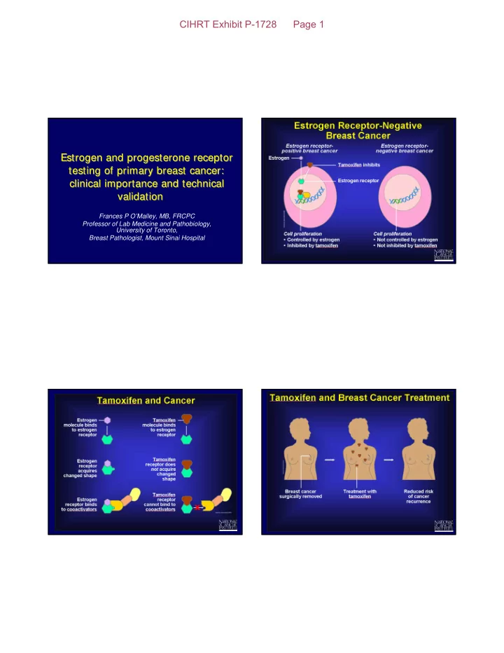
Estrogen and progesterone receptor testing of primary breast cancer: clinical importance and technical validation Estrogen and progesterone receptor testing of primary breast cancer: clinical importance and technical validation
Frances P O’Malley, MB, FRCPC Professor of Lab Medicine and Pathobiology, University of Toronto, Breast Pathologist, Mount Sinai Hospital