Epilepsy Vikram Rao, MD, PhD NeuroPace, Inc.: Consultant Diagnosis - - PowerPoint PPT Presentation

epilepsy
SMART_READER_LITE
LIVE PREVIEW

Epilepsy Vikram Rao, MD, PhD NeuroPace, Inc.: Consultant Diagnosis - - PowerPoint PPT Presentation

2/14/2019 Disclosures Epilepsy Vikram Rao, MD, PhD NeuroPace, Inc.: Consultant Diagnosis & Management Upsher Smith Laboratories: Served on Scientific Advisory Board Switch Bio, Inc.: Served on Scientific Advisory Board 2019 Neurona,


slide-1
SLIDE 1

2/14/2019 1

Epilepsy Diagnosis & Management 2019

Susannah B. Cornes, MD Associate Professor of Clinical Neurology Vikram R. Rao, MD, PhD Assistant Professor of Clinical Neurology

Disclosures

Vikram Rao, MD, PhD NeuroPace, Inc.: Consultant Upsher Smith Laboratories: Served on Scientific Advisory Board Switch Bio, Inc.: Served on Scientific Advisory Board Neurona, Inc.: Consultant Susannah Cornes, MD None

  • Seizures: paroxysmal, stereotyped spells of

altered movement, sensation, experience, and/or consciousness resulting from excessive brain electrical activity

Institute of Medicine Report (2012); www.cureepilepsy.org; www.cdc.gov; Zack and Kobau, MMWR Morb Mortal Wkly Rep (2017) 66:31

Epilepsy

  • Epilepsy: recurrent, unprovoked seizures
  • 4th most common neurological disorder (migraine, stroke, AD)
  • ∼1% of the population (3.4 million in U.S.)
  • 1 in 26 people will develop epilepsy in their lifetime
  • Etiology: stroke, tumor, trauma, infection, genetic, metabolic,

developmental, neurodegenerative, cryptogenic, …

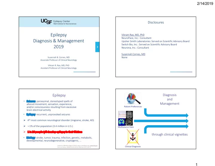
Patient Preferences Clinical Diagnosis Multisource Data

Diagnosis and Management through clinical vignettes

slide-2
SLIDE 2

2/14/2019 2

CASE 1: 33 RHM h/o HTN, HL p/w 6-months of stereotyped spells Internal head sensaon → R face numb and weak, dysarthria → R hand/arm numb and weak, tremoring movements → R leg weak; no loss of awareness Duration: 4–8 min, Frequency: 2-3x/month

Workup Head CT, brain MRI, TTE, Holter, carotid U/S, routine EEG all normal Lupus anticoagulant positive; started on lovenox → warfarin Spells persisted despite therapeutic INR Focal seizures suspected, Keppra started Trileptal added with minimal improvement Referred to UCSF for refractory epilepsy

Audience Response System At this point, the MOST likely diagnosis is:

  • A. Jacksonian epilepsy
  • B. Cardioembolic TIA
  • C. Complex migraine
  • D. Call-Fleming syndrome
  • E. Other vascular phenomenon

6 Confidential – 9/21/15

Jacksonian epilepsy Cardioembolic TIA Complex migraine Call-Fleming syndrome Other vascular phenom...

67% 4% 7% 4% 18%

UCSF Evaluation

Red flags:

  • spells involve mostly negative symptoms (numbness, weakness)
  • right arm shaking described as oscillatory (not clonic)
  • relatively long duration (>4 min)
  • too stereotyped for cardioembolic process
  • no response to two antiepileptic drugs

Additional history:

  • >90% of events occurred while in a hot shower or upon standing

Further workup:

  • Brain MRI/A ordered

UCSF Evaluation

Brain MRI/A: High-grade left M1 stenosis, no infarcts CTA: confirmed 70% left M1 stenosis CTP: left MCA territory ischemia Tapered off Keppra, Trileptal; started on aspirin, statin, permissive HTN; referred to UCSF Neurovascular MRA CTA CTP

slide-3
SLIDE 3

2/14/2019 3

UCSF Treatment

Wingspan stent to left M1 Aspirin + Plavix Spells resolved completely

MRA

CASE 2: 54 RHF with 28 years of spells of R hemibody paresthesias Indescribable aura, electrical head sensation that shoots down right hemibody, occ. falls, no loss of awareness Duration: 10 sec, Frequency: catamenial, clusters up to 100/day; Triggers: stress, alcohol, sleep deprivation

Workup Head CT, brain MRI, sleep-deprived EEG, carotid U/S, TCD all normal Improved spell frequency on Dilantin, Keppra added without benefit Diagnosed with “psychosomatic” hemisensory syndrome Advised to reduce stress, improve diet/exercise, ensure good sleep She presented to UCSF for a second opinion

33/34 patients with extensive workup negative, 1 patient with corona radiata stroke

Audience Response System At this point, the MOST likely diagnosis is:

  • A. Multiple sclerosis
  • B. Psychogenic non-epileptic

spells

  • C. Focal sensory seizures
  • D. TIA
  • E. Catamenial migraine

13 Confidential – 9/21/15

Multiple sclerosis Psychogenic non-epilept.. Focal sensory seizures TIA Catamenial migraine

10% 7% 29% 0% 54%

UCSF Evaluation

Red flags:

  • consistent aura before symptoms
  • falls with some episodes
  • no psychiatric risk factors for non-epileptic spells
  • spells improved on Dilantin
  • reported triggers (stress, alcohol, sleep deprivation) are common

for seizures Additional history:

  • prior EEGs had not been done during symptomatic periods

Further workup:

  • 48-hour ambulatory EEG ordered, timing to be based on

catamenial variation in spells

slide-4
SLIDE 4

2/14/2019 4

UCSF Evaluation

Ambulatory EEG Frequent left parietal spikes No clear ictal correlate to patient’s symptoms Auras can be EEG-negative Symptoms presumed epileptic Seizure-free on oxcarbazepine

CASE 3: 52-year old man h/o OSA, two cousins with epilepsy Onset of spells at age 16 Hypermotor nocturnal events involving brief (<15 sec) tonic extensor posturing and flailing limb movements; Duration: <15 sec; Frequency: multiple times every night

Workup Brain MRI reportedly normal Routine EEG: negative Sleep study: ?REM behavior disorder, no seizure activity Epilepsy considered possible, treated with 3 antiepileptic drugs Referred to UCSF for pre-surgical evaluation

Localizing seizures in the brain

Scalp EEG 3T MRI PET MEG SPECT Stereo-EEG Grid electrodes Stimulation mapping Wada

UCSF Evaluation

Video-EEG: no interictal spikes, 3 seizures without EEG correlate PET-CT: negative Neuropsychology: non-lateralizing patterns of deficits 3T Brain MRI:

Transmantle focal cortical dysplasia (likely FCD IIb) Highly epileptogenic lesion High likelihood of overlap with eloquent cortex

slide-5
SLIDE 5

2/14/2019 5

Audience Response System At this point, the MOST likely appropriate next step ?:

  • A. Lesionectomy
  • B. Intracranial EEG
  • C. Implant responsive

neurostimulator

  • D. Implant vagus nerve stimulator
  • E. Cognitive behavioral therapy

20 Confidential – 9/21/15

Lesionectomy Intracranial EEG Implant responsive neur... Implant vagus nerve stim... Cognitive behavioral the...

3% 51% 0% 7% 39%

UCSF Evaluation

Intracranial EEG: 122 subdural electrodes (grid/strips/depths)

  • Seizure onset zone localized to the dysplasia and adjacent

interhemispheric cortex

  • Electrical stimulation mapping revealed overlap with

eloquent (leg) motor area

  • Patient unwilling to accept treatment-related neurological

deficits

Advanced treatments for seizures

Resection Laser Devices

Electrodes

  • Record from seizure focus/foci
  • Stimulate to reduce seizures

Leads

  • Strips or depths

Responsive Neurostimulator

  • Cranially implanted
  • Electronics and battery
slide-6
SLIDE 6

2/14/2019 6

https://...

Chronic electrocorticography

RNS leads Electrocorticogram of seizure One year after responsive stimulation was enabled, patient is completely seizure-free

Responsive neurostimulation

CASE 4: 28 RHF with onset of epilepsy at age 12 Right leg tingling and ascending numbness, right leg stiffens and jerks, falls, no loss of awareness Duration: 60 sec; Frequency: 3x/week, secondarily generalizes to convulsion 1x/month

Workup 3T Brain MRI: normal Video-EEG:

  • Interictal: left centro-parietal spikes
  • Ictal: left centro-parietal seizure onset

PET-CT: dorsal-medial parietal hypometabolism Treated with 3 antiepileptic drugs Intracranial EEG for precise seizure localization

PET EEG Source Localization

dorsal view inferior view

Intracranial Monitoring

  • Seizure onset from centroparietal region
  • Stimulation mapping revealed overlap with eloquent (leg)

sensorimotor cortex

  • Patient willing to risk neurological defict for an intervention

with highest chance of seizure freedom

slide-7
SLIDE 7

2/14/2019 7

Audience Response System Which one of the following is generally associated with the HIGHEST rate of seizure freedom?:

  • A. Thalamic deep brain stimulation
  • B. Addition of a fourth antiepileptic

drug

  • C. Responsive neurostimulation
  • D. Resective surgery
  • E. Multiple subpial transections

29 Confidential – 9/21/15 Thalamic deep brain stim... Addition of a fourth anti... Responsive neurostimula... Resective surgery Multiple subpial transect...

5% 0% 5% 84% 5%

Surgical pathology: Focal cortical dysplasia IIb Clinical outcome: Seizure-free x 9 months, minimal deficits

Resective surgery

Tailored neocortical resection

CASE 5: 21yo W p/w new onset shaking spells 2w s/p cesarian in the setting of severe pre-eclampsia Post-partum depression, anxiety, behavioral changes Placed on risperidone for post-partum psychosis Interactive between frequent episodes of shaking

Work-Up: CT, MRI normal; CTX, Vanc, Flagyl, Acyclovir, Dilantin; LP normal Additional history: Keppra added; noted to be increasingly sedated so Dilantin weaned. Risperidone stopped due to abnormal buccal movements. Transferred to UCSF for Video-monitoring to diagnose spells.

Audience Response System At this point, the MOST likely diagnosis is:

  • A. Eclampsia
  • B. Psychogenic nonepileptic

spells

  • C. Seizure provoked by

antipsychotics

  • D. Paraneoplastic encephalitis
  • E. New onset epilepsy

33 Confidential – 9/21/15

Eclampsia Psychogenic nonepileptic... Seizure provoked by anti... Paraneoplastic encephalitis New onset epilepsy

8% 37% 22% 24% 10%

slide-8
SLIDE 8

2/14/2019 8

UCSF Evaluation

Red flags:

  • spells of twitching refractory to two medications
  • additional nonstereotyped dyskinetic movements of the face
  • increasingly encephalopathic
  • onset occurred >1 week post-partum

Additional history:

  • exam on arrival was unresponsive

to voice or noxious stimulation

  • repeat MRI with B hippocampal FLAIR

3T MRI

UCSF Evaluation

Frequent Motor Seizures Interictal Background Further work-up:

  • EEG demonstrated frequent

motor seizures

  • interictal background was

diffuse slow with excess beta

  • repeat LP bland, sent for

autoimmune encephalitis panel

UCSF Evaluation and Treatment

Further Work-up:

  • NMDA-R Ab +

Ongoing Treatment:

  • s/p removal of teratoma
  • s/p multiple AEDs
  • IVIG, IVSM, Rituximab
  • remains on CLZ, LEV
  • no e/o seizure but with

persistent encephalopathy after NMDA mediated syndrome

Schmidt et al. Neurology. 2012 Sept 11;79(11):1094-100

Diffuse slowing, excess beta = Extreme delta brushes Pelvic US: 0.7cm echogenic focus R ovary

CASE 6: 22yo RH M, +FH FS, IEP, onset 8yo GTC on awakening EEG with generalized spike and wave Every 3-12 mo → 4-12/mo by 18y No aura, ictal cry, BUE flexion, gen shaking 3 diurnal; 1 during basketball (kept dribbling); no aura.

Work-Up:

MEDs: LEV, VPA, ZNS, LRZ VET Interictal: GSW, polyspike VET Ictal: 3 GTC with L emphasis. Ext RUE prior to LUE. MRI: Normal

Other history:

Changed to LTG, VPA Noted new daily myoclonus Clusters of nausea, feverishness, goosebumps, x 3d / 1mo.

Generalized spike and wave Generalized onset

slide-9
SLIDE 9

2/14/2019 9

Audience Response System At this point, the BEST next step is:

  • A. Discontinue Lamotrigine
  • B. Readmit for additional Video-

EEG

  • C. Add a benzodiazepine
  • D. Evaluation for IBD
  • E. Repeat MRI

39 Confidential – 9/21/15

Discontinue Lamotrigine Readmit for additional V... Add a benzodiazepine Evaluation for IBD Repeat MRI

17% 52% 23% 5% 3%

UCSF Evaluation

Red flags:

  • unilateral automatic movements prior to one seizure
  • paroxysmal piloerection and autonomic sx’s c/w temporal aura
  • refractoriness
  • aura history not reliable for seizures from sleep

L TIRDA L anterotemporal sharps L FIAS

UCSF Evaluation

Additional history:

  • addional AED trials: TPX, CLN, CLB; LCS, OXC → myoclonus
  • MSI recorded focal L temporal sz; MRI normal; PET symmetric
  • iEEG recording L temporal/frontal
  • hippocampal herald spike → OFC, temp grid; nausea sensaon
  • L temporal lobectomy
  • Path c/w MTS
  • 3 years seizure free

CASE 7: 42yo W onset 8yo symptomatic seizures Resection L frontal oligodendroglioma 10yo Recurrence 11yo with refractory aura of pressure feeling in her head 2-3 x /week Occasionally → eyes pulling to the right

Work-Up: Prior AEDS: VPA, PB, CBZ Current: LTG, CLB Admitted for video EEG: L frontocentral sharps…

slide-10
SLIDE 10

2/14/2019 10

UCSF Evaluation

MRI MSI

Additional history:

  • pressure sensation aura had no EEG correlate
  • 1 nonepileptic event, falling back against bed

unresponsive

Source Localization

Audience Response System What is your next BEST step:

  • A. Refer for CBT for PNES
  • B. ECOG guided perilesional

resection

  • C. Add treatment for headache
  • D. Phase 2 intracranial recording
  • E. Repeat scalp video-EEG

telemetry

45 Confidential – 9/21/15

Refer for CBT for PNES ECOG guided perilesional... Add treatment for head... Phase 2 intracranial rec... Repeat scalp video-EEG t...

11% 10% 15% 62% 2%

UCSF Evaluation

Movie

Additional history:

  • interictal ECOG sharps at C6, C7 inferior and

posterior to lesion

  • motor mapping demonstrated expected

findings along precentral gyrus

  • aura mapping demonstrating FEF within

precentral gyrus associated with seizure feeling

UCSF Treatment

Additional history:

  • region of ECOG sharps

included in resection

  • RNS placed overlying

region of aura within precentral sulcus

  • seizure free including

pressure sensation

slide-11
SLIDE 11

2/14/2019 11

CASE 8: 33yo RHF with hx IEP, childhood onset, failed 7 AEDs Fumbling with hands, lip smacking, L head turn, 4/mo L posterior quadrant seizures by vEEG Auditory and confrontational naming deficits Concordant MEG/MSI, PET, MRI (L inferior temporal FCD)

MRI: structural lesion (FCD) EEG: focal left temporo-

  • ccipital seizure onset

MSI: spike cluster basal posterior L temporal

UCSF Evaluation

iEEG: Ictal onset over basal mini-grid

Further work-up:

  • Unilateral iEEG
  • Onset basal temporal
  • Language mapping
  • Basal temporal resection

UCSF Evaluation

Additional history:

  • recurrence of daily seizures in the first post-operative week

Post-op MRI of iEEG-guided resection

Audience Response System At this point, the management plan MOST likely to achieve seizure freedom is:

  • A. Implant responsive

neurostimulator

  • B. Readmit for additional Video-

EEG

  • C. Add benzodiazepine
  • D. Extend lesionectomy
  • E. Implant vagus nerve stimulator

52 Confidential – 9/21/15

I m p l a n t r e s p

  • n

s i v e n e u r . . . R e a d m i t f

  • r

a d d i t i

  • n

a l V . . . A d d b e n z

  • d

i a z e p i n e E x t e n d l e s i

  • n

e c t

  • m

y I m p l a n t v a g u s n e r v e s t i m . . .

33% 20% 4% 43% 0%

slide-12
SLIDE 12

2/14/2019 12

UCSF Treatment

Additional history:

  • repeat resection 2.5 months after first surgery
  • path moderate gliosis only
  • seizure free for >2 year; single seizure during nonadherence
  • returned to work as special education teacher

Impact on seizure freedom after repeat surgery Odds Ratio

Congruent electrophysiology 3.6 (1.6 – 8.2)* Lesional versus nonlesional pathology 3.2 (1.9 – 5.3)* Surgical limitations as cause for initial surgery failure 2.6 (1.3 – 5.3)* Temporal lobe resection 1.5 (0.8 - 3.0) Abnormal pre-operative MRI 1.9 (0.6 – 5.4) Prior iEEG 0.4 (0.2 – 0.9)

N=782; Engel 1 47% overall → 58% with 1 predictor. (Krucoff et al. Epilepsia. 2017; 58(12):2133-2142)

Take-Home Points

  • Epilepsy is a clinical diagnosis.
  • Be vigilant for red flags that promote diagnostic

error (framing, anchoring, premature closure).

  • Treatment decision should account for patient’s

goals and risk tolerance.

  • Potential for seizure freedom more likely with

multisource concordant data.

  • There is often more than one path to a good
  • utcome.

Acknowledgements

Neurology

Dan Lowenstein, MD Paul Garcia, MD Robert Knowlton, MD Heidi Kirsch, MD Tina Shih, MD Susannah Cornes, MD Manu Hegde, MD, PhD Vikram Rao, MD, PhD Mercedes Paredes, MD, PhD Joseph Sullivan, MD Roberta Cilio, MD, PhD Nilika Singhal, MD Adam Numis, MD

Neurosurgery

Edward Chang, MD Kurtis Auguste, MD

Nursing

Maritza Lopez, RN Kseniya Mandic, NP Mariann Ward, NP

Neuropsychology

Brandon Kopald, PhD Brianna Paul, PhD

Neurodiagnostics

Mary Betinis, R. EEG T. Tommy Thompson, R. EEG T.

Fellows

Jon Kleen, MD, PhD Brandy Ma, MD Rawan Daghistani, MD