Early Vitamin K Defjciency- A Rare Presentation Merchant R 1 , Doctor - - PDF document

early vitamin k defjciency a rare presentation
SMART_READER_LITE
LIVE PREVIEW

Early Vitamin K Defjciency- A Rare Presentation Merchant R 1 , Doctor - - PDF document

Open Access Journal of Radiology and Diagnostjc Methods REVIEW ARTICLE Early Vitamin K Defjciency- A Rare Presentation Merchant R 1 , Doctor P 2 , Kulkarni S 1 , Choudhari A 1 and Pandey AK 3* 1 Consultant Department of Pediatrics, Nanavati Super


slide-1
SLIDE 1

Early Vitamin K Defjciency- A Rare Presentation

Merchant R1, Doctor P2, Kulkarni S1, Choudhari A1 and Pandey AK3*

1Consultant Department of Pediatrics, Nanavati Super Speciality Hospital, Mumbai, India 2Pediatric Department, Nanavati Super Speciality Hospital, Mumbai, India 3Medical Intern, Nanavati Super Speciality Hospital, Mumbai, India

REVIEW ARTICLE Open Access

Volume 2 | Issue 1

ScholArena | www.scholarena.com

Journal of Radiology and Diagnostjc Methods

Vitamin k is an anti-hemorrhagic factor that is needed for the synthesis of functional forms of factor II, VII, IX, and X in the liver. Once activated in the blood, they become available to take part in the coagulation process, a complex series of events that results in the conversion of fjbrinogen to fjbrin and the formation of a hemostatic plug. Tie consequence of vitamin k defjciency results into a hypocoagulable state, however, the hemostatic system can function adequately at low-factor concentrations but as the defjciency progresses, a point will reach when the procoagulatory mechanisms fail and bleeding occurs. Newborns have only 20–50% of adult coagulation activity. Lack of vitamin K administration at birth, exclusive breastfeeding, chronic diarrhea and prolonged use of antibiotics make them more prone to Vitamin K defjciency bleeding (VKDB) [1]. VKDB is a well-known entity and presents in 3 difgerent clinical forms: early, classical and late. Classical VKDB develops mainly as gastrointestinal hemorrhage from day 2 to 7, and late VKDB develops as mainly intracranial hemorrhage from 2 weeks to 6 months. Compared with classical and late VKDB, early neonatal VKDB causes mainly internal hemorrhage within 24 h afuer birth along with a higher mortality rate compared to classical and late VKDB [3].

Introduction Case Report

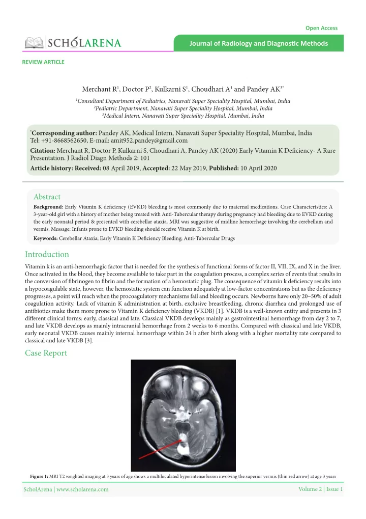
Background: Early Vitamin K defjciency (EVKD) bleeding is most commonly due to maternal medications. Case Characteristics: A 3-year-old girl with a history of mother being treated with Anti-Tubercular therapy during pregnancy had bleeding due to EVKD during the early neonatal period & presented with cerebellar ataxia. MRI was suggestive of midline hemorrhage involving the cerebellum and

  • vermis. Message: Infants prone to EVKD bleeding should receive Vitamin K at birth.

Abstract

Keywords: Cerebellar Ataxia; Early Vitamin K Defjciency Bleeding; Anti-Tubercular Drugs

*Corresponding author: Pandey AK, Medical Intern, Nanavati Super Speciality Hospital, Mumbai, India

Tel: +91-8668562650, E-mail: amit952.pandey@gmail.com Citation: Merchant R, Doctor P, Kulkarni S, Choudhari A, Pandey AK (2020) Early Vitamin K Defjciency- A Rare

  • Presentation. J Radiol Diagn Methods 2: 101

Article history: Received: 08 April 2019, Accepted: 22 May 2019, Published: 10 April 2020

Figure 1: MRI T2 weighted imaging at 3 years of age shows a multiloculated hyperintense lesion involving the superior vermis (thin red arrow) at age 3 years

slide-2
SLIDE 2

J Radiol Diagn Methods

2

Volume 2 | Issue 1

ScholArena | www.scholarena.com

Discussion

Her mother was diagnosed with pulmonary tuberculosis in the third trimester and was started on anti-tuberculosis treatment with Isoniazid, Rifampicin, and Ethambutol. Antenatal ultrasounds at 38 weeks gestation were normal with no intracranial pathology noted. Tie child was born full term by lower segment cesarean section, cried immediately afuer birth but developed seizures and required mechanical ventilation within 12 hrs of delivery. Investigations revealed prolonged prothrombin time (52s), partial thromboplastin time (60s) and INR of 4.2 which corrected to normal afuer injecting 1 mg intramuscular (IM) Vitamin K. Ultrasound of the brain performed on day 5 of life suggested a hypoechoic lesion with mild peripheral vascularity in the cerebellum measuring 1.5 cm in all dimensions. An MRI brain on day 5 confjrmed midline hemorrhage involving the cerebellum and vermis measuring 2.1 × 1.6 × 1.2 cms in dimension (Figure 2). Tie child was treated with phenytoin and phenobarbital for seizure control. Transplacental transfer of vitamin K is very limited during pregnancy, as is the storage of vitamin K in the neonatal liver, all of which makes the newborn infant uniquely vulnerable to hemorrhagic disorders unless exogenous vitamin K is given immediately afuer birth [2]. Classically, vitamin K defjciency bleeding is observed in infants who have not received prophylactic vitamin K at birth, with an incidence ranging from 0.25 to 1.7 cases per 100 births. Today, it is the standard of care around the world to provide vitamin K supplementation to the newborn with a single intramuscular dose (range, 0.5–1 mg) within 1 hour of birth. VKDB can be due to idiopathic cause or secondary causes. In secondary VKDB, there is an underlying cause, usually an undiagnosed disease such as hereditary hepatobiliary/ malabsorptive disease (e.g: - biliary atresia, alpha-1-antitrypsin defjciency, cystic fjbrosis) or the efgect of drugs that have been given to mother or infant. Bleeding commonly occurs in the umbilicus, gastrointestinal (GI) tract, skin, epistaxis, surgical sites (ie, circumcision) and, uncommonly, in the brain [4]. Early VKDB is almost always associated with maternal medications that interfere with vitamin K metabolism such as anticonvulsants (phenytoin, barbiturates, carbamazepine), antitubercular drugs (rifampin and isoniazid), antibiotics (cephalosporins) and vitamin K antagonists (warfarin and coumarin) [5]. Tie incidence of early VKDB in neonates of mothers taking these medications without vitamin K supplementation varies from 6% to 12% [5]. Although Intracranial Haemorrhage occurs most ofuen in late VKDB (50-75%), it may occur in 20-25% of cases of early VKDB [6]. Common signs and symptoms in VKDB newborns with ICH are convulsions (80%), feeding intolerance and poor sucking (50%), irritability (40%) vomiting (47%) and fever (40%) [7]. Tie prognosis in patients with ICH due to VKDB depends

  • n early diagnosis, rapid and adequate corrections of coagulation defect, and the general condition of the patient, including factors

such as the presence of anemia and rapid surgical intervention [3]. Fresh frozen plasma (FFP) can be given to patients with normal hemoglobin level, with severe ICH as it contains all blood-clotting factors. Intravenous injection of vitamin k in the treatment of VKDB results in signifjcant reversal of the hemostatic defect within minutes and its prompt use may obviate the need to administer plasma products [1]. In a study done by Zidan et al., despite early surgical evaluation, the mortality rate was high along with neurological complication. We present a unique case of a 3-year-old child who presented with seizures during the A 3-year-old girl fjrst by birth order, born of a non-consanguineous marriage was noticed to have diffjculty in balancing while walking and slurred speech since 2 years of age. Tie child started sitting at 12 months, months suggesting a motor developmental

  • delay. On examination, she had staccato speech, hypotonia, dysmetria, and ataxic gait. Magnetic resonance imaging (MRI) brain

revealed multiloculated fmuid intensity cystic areas involving the superior cerebellar vermis and hippocampus with hemosiderin staining the wall of the cysts suggestive of a sequela of hemorrhage (Figure 1).

Figure 2: MRI T1 weighted imaging on day 5 of age showed a midline hemorrhage involving the cerebellum and vermis measuring 2.1 × 1.6 × 1.2 cms in dimension (thick red arrow)

slide-3
SLIDE 3

J Radiol Diagn Methods

3

Volume 2 | Issue 1

ScholArena | www.scholarena.com early neonatal period secondary to early VKDB in the cerebellum and later developed cerebellar ataxia and motor delay due to a remnant cyst formed in the superior cerebellar vermis. Infants predisposed to manifest early VKDB as evidenced by maternal intake of medications mentioned above should receive vitamin K, 1 mg IM at birth and they may be delivered by cesarean section to avoid the trauma of vaginal delivery [8,9]. RM, PD, SK, AP made substantial contributions to the design of the work by going through the history and physical of the patients along with the interpretation of past medical records. All the radiological interpretations were done by AC. Tie drafu was prepared by PD, AP whereas RM, AC and SK revised it for important intellectual content. Afuer approval from all the authors, the case report was ready for fjnal submission. Tie complete case report was prepared at the Department of Pediatrics, Nanavati super specialty hospital. Tie authors have none to declare.

References Contribution of Authors Confmict of Interest and Source of Support

  • 1. Shearer MJ (2009) Vitamin K defjciency bleeding (VKDB) in early infancy. Blood Rev 23: 49-59.
  • 4. Pichler E, Pichler L (2008) Tie neonatal coagulation system and the vitamin K defjciency bleeding - a mini review. Wien Med Wochenschr. 158: 385-95.
  • 5. Lippi G, Franchini M (2011) Vitamin K in neonates: facts and myths. Blood Transfus 9: 4-9.
  • 6. Takahashi D, Shirahata A, Itoh S, Takahashi Y, Nishiguchi T et al. (2011) Vitamin K prophylaxis and late vitamin K defjciency bleeding in infants: fjfuh nationwide

survey in Japan. Pediatr Int 53: 897-901

  • 7. Yilmaz C, Yuca SA, Yilmaz N, Bektas MS, Caksen H (2009) Intracranial hemorrhage due to vitamin K defjciency in infants: a clinical study. Int J Neurosci 119:

2250-6.

  • 8. Sankar MJ, Chandrasekaran A, Kumar P, Tiukral A, Agarwal R, et al. (2016) Vitamin K prophylaxis for prevention of vitamin K defjciency bleeding: a systematic
  • review. J Perinatol 36: 29-35.
  • 9. McNinch A (2010) Vitamin K defjciency bleeding: early history and recent trends in the United Kingdom. Early Hum Dev 1: 63-5.
  • 2. Elalfy MS, Elagouza IA, Ibrahim FA, AbdElmessieh SK, Gadallah M (2014) Intracranial haemorrhage is linked to late onset vitamin K defjciency in infants aged

2-24 weeks. Acta Paediatr 103: e273-6.

  • 3. Zidan AS, Abdel-Hady H (2011) Surgical evacuation of neonatal intracranial hemorrhage due to vitamin K defjciency bleeding. J Neurosurg Pediatr 7: 295-9.