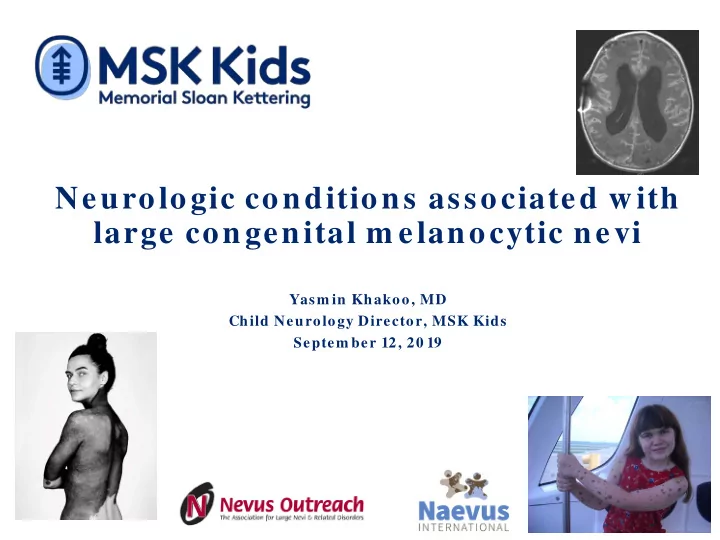
SLIDE 1 Neurologic conditions associated with large congenital m elanocytic nevi
Yasm in Khakoo, MD Child Neurology Director, MSK Kids Septem ber 12, 20 19
SLIDE 2 Disclosures
Provides funding for MSK NCM database Reim burses for m eeting/ travel related expenses
2
SLIDE 3
Outline
Definition History Criteria Incidence and epidemiology Embryology Mouse models/ genetics Neuropathology Screening recommendations Neurologic complications Therapeutic targets
SLIDE 4 Photo cred: Brock Elbank and Caring Matters Now
SLIDE 5
Definition
A rare neurocutaneous syndrome defined by the presence of large and/ or multiple congenital cutaneous nevi and melanocytes in the CNS
SLIDE 6
SLIDE 7 A B
MRI brain
Coronal post contrast Axial T1 FLAIR
SLIDE 8 Slutsky et al, Sem in Cut Med and Surg 2010
SLIDE 9
One of our recent patients
SLIDE 10 History
1861: Viennese pathologist Karl Rokitansky described autopsy findings of a 14 yo girl with developmental delay and nevi
(Translation: “A remarkable case of a pigmented nevus with extensive leptomeningeal pigmentation”)
SLIDE 11
History continued
1948: Van Bogaert named the syndrome neurocutaneous melanosis (NCM) 1991: Kadonaga and Frieden outlined criteria 2000: Nevus Outreach, Inc registry formed 2005: Marghoob: NCM in pts with LCMN: 7% 2007: Bauer: NRAS mutations in LCMN 2012: Shakhova: Mouse model 2012: Kinsler estimated 18% of LCMN have brain lesions 2013: Kinsler detects NRAS mutations in brain lesions 2018: Naevus International formed
SLIDE 12 Photo cred: Brock Elbank and Caring Matters Now
SLIDE 13 Criteria
Presence of large (>20 cm) and/ or multiple (>3) congenital melanocytic nevi (CMN) with meningeal melanosis or melanoma;
Must distinguish between metastatic melanoma and primary
1991, J Am Acad Derm
SLIDE 14 Incidence/epidemiology
A mostly sporadic condition (one report of 2 siblings) LCMN: 1/ 20,000 brain 1/ 200,000 M=F Age was ~ 3 but with MRI, earlier diagnosis Majority of patients who will become symptomatic do so by 2 years; 70% by 5 yrs Reports of patients who become symptomatic in 2nd
Use of MRI and other radiographic techniques may increase the number of patients identified
SLIDE 15 Embryology
Melanocytes develop from neural crest and migrate throughout the body including the covering of the brain (leptomeninges) Nevi: melanocytes which arrest along the path NCM may be a marker for abnormal neuronal migration
Wolff et al: E11.5 mouse LacZ staining for melanoblasts
SLIDE 16 2007, J Invest Derm at
Mutation found in codon 61 of the NRAS gene
SLIDE 17 2012 Nat Cell Biol
SOX10 important in neural crest development SOX10 highly expressed in LCMN and melanoma NRAS Q61 controls the expression of SOX10
SLIDE 18 2013 J Invest Derm
Twelve of 15 patients tested had NRAS mutations in affected skin and neural tissue Normal tissue and blood were normal Ten had a Q61K mutation while 2 had Q61R LOH was seen in 2 patients who developed cutaneous melanoma We identified same mutations in 2 patients
SLIDE 19
Neuropathology: Gross
SLIDE 20 Low and high power views of the cortex infiltrated with pigmented cells
Neuropathology: microscopic
Leptomeningeal and sulcal melanocytosis (courtesy M. Reyes-Mugica)
CSF with nevo-melanocytes
SLIDE 21 Patients with posterior midline LCMN had moderate risk Patients with 20 or more satellite nevi are at high risk
(Arch Derm at 2004)
SLIDE 22 Photo cred: Brock Elbank and Caring Matters Now
SLIDE 23
Neurologic complications
Some children with brain lesions have NO neurologic complications Conversely, some patients with LCMN and normal MRI have neurologic complications Hydrocephalus Seizures Cranial/ spinal nerve dysfunction Developmental delay Spinal cord compression/ tethered cord
SLIDE 24
Temporal lobe melanocytosis and Dandy- Walker cyst
SLIDE 25 Hydrocephalus
Decreased outflow either communicating or
Axial T1 post contrast MRI: diffuse leptomeningeal enhancement
SLIDE 26 Hydrocephalus: treatment
Either a ventriculo-peritoneal (VP) shunt or endoscopic third ventriculostomy (ETV) If protein or cells in spinal fluid is high, shunt
New programmable shunts may improve
SLIDE 27
Hydrocephalus: signs and symptoms
Headaches (irritability, head banging in pre- verbal child) Enlarging head circumference Morning nausea and vomiting Limited upgaze (Sun setting eyes) Diplopia (CN VI palsy) Lower extremity spasticity
SLIDE 28 Seizures
- Causes of seizures in patients with LCMN
- Abnorm al neuronal m igration
- Typically have partial seizures;
- 2 infants presented with infantile spasms
- Treatment
- Anticonvulsants
- Surgery if a focus can be identified
SLIDE 29
Cranial nerve symptoms/signs
May have diminished hearing because of melanocytic deposits on auditory nerves
Screening audiogram recommended for all patients Sign language
All patients should have baseline eye exam
SLIDE 30
Developmental/behavioral issues
May result from chronic neurologic conditions May also be due to psychological effects of Early intervention for all children <3 yrs May need resource room as child gets older Certain anticonvulsants may be helpful for treating behavioral issues as well
SLIDE 31
Spinal nerve root and cord compression
Cord compression from nodules or tethered cord Delayed motor milestones or toe walking (myelopathy) Delay in toilet training Back pain Treatment of tethered cord: surgical release Cord compression: symptomatic: steroids, surgical decompression
SLIDE 32
Tethered cord with associated syrinx
SLIDE 33
Spinal arachnoid cyst
SLIDE 34 Photo cred: Brock Elbank and Caring Matters Now
SLIDE 35 IRB approved Methods: Chart review 2003-2010 Results
- 14 patients LCMN/ NCM identified
- All had MRI brain, spine
- 8/ 14 patients alive at a median age of 3 years
- 6 had diffuse leptomeningeal enhancement
- 3 had spinal arachnoid cysts
- 1 had a benign cervical spindle cell tumor
2012 Dev Med Child
SLIDE 36
Results
5/ 14: Asymptomatic median age 48 mos 7/ 14: Seizures; 5 had sz as initial neurologic symptom 11 were normal or had mild developmental delay
3 had moderate –severe developmental delay 2 had diffuse leptomeningeal enhancement
2/ 14 had symptomatic hydrocephalus
Median age 16.2 mos (birth-8yrs)
4/ 5 with diffuse leptomeningeal enhancement had primary CNS melanoma
SLIDE 37
Neurologic summary
Children with mild to moderate NCM can live with some neurologic deficits Presence of diffuse leptomeningeal disease portends poor prognosis High incidence of spinal cystic malformations Imaging of the entire neuraxis should be performed in all children with large congenital melanocytic nevi, ideally before 4 months of age
SLIDE 38
Screening recommendations
Patients with >20 satellite nevi and/ or LCMN >20 especially 40c cm baseline MRI of the brain and spine with and without contrast before age 4 months Risk of anesthesia needs to be considered
SLIDE 39
How many MRIs?
If initial MRI normal and child neurologically normal, no further imaging needed If initial MRI positive for brain lesions child should be followed closely by child neurologist If child continues to be neurologically asymptomatic, repeat MRI not indicated If any clinical change MRI should be repeated
SLIDE 40
Food for thought
One patient had “disappearance” of brain lesions Isolated reports of patients developing vitiligo who then have regression of brain lesions
SLIDE 41 Photo cred: Brock Elbank and Caring Matters Now
SLIDE 42 CNS melanoma CNS melanosis
21 12 (36%) (64%)
Courtesy Ashfaq Marghoob
NCM→Melanoma? Symptomatic NCM: 33 cases reviewed
SLIDE 43 CNS melanoma treatment options
Interferon alpha and IL-2 have not been effective Temozolomide oral chemotherapy may help prolong survival Platinum based IV chemotherapy may also prolong survival Ipilumimab Intrathecal radio-immunotherapy may be more specific Non malignant brain lesions: no binding to 3F8
Poor CSF flow when LMD is widespread
SLIDE 44 IT radiolabeled antibody: how does it work?
3F8 or 8H9 antibody selectively binds to tumor cells of neural crest origin (e.g. melanoma, medulloblastoma, neuroblastoma)
Courtesy of Dr. Kim Kram er
SLIDE 45 Other targets : NRAS pathway
Somatic NRAS mutations identified in patients with LCMN (Papp et al, 1999) Brain lesions also contains NRAS mutations
(Kinsler et al, 2013)
LCMN and brain lesions (esp with nodular features) may also contain BRAF mutations (Salgado et al, 2015) We tested CNS tissue from two patients (one temporal lobe and one SC) and found same mutation
SLIDE 46 Pharmacologic inhibition of ERK signaling
GRB2 SOS
NRAS
NF1 MEK2 MEK1 ERK1/2 BRAF
RAF inhibitors: Vem urafenib Dabrafenib *Only BRAF V60 0 E tum ors MEK inhibitors: PD0 32590 1 AZD6244 (selum etinib) GSK1120 212 (tram etinib) Binim etinib
Courtesy of C. Pratilas
SLIDE 48 Randi Silver PhD at Weill Cornell studies wound healing Patients with LCMN have increased numbers of mast cells in tissue We tested 2 CNS samples but did not detect presence of mast cells
2014 Ped Dev Pathol
SLIDE 49 Nevospheres were isolated from skin, brain and spinal cord of patients with LCMN and brain lesions All tissues harbored NRAS mutations Vemurafenib (BRAF inhibitor) was not effective MEK inhibitor only partially inhibited PI3K and mTOR inhibitors seemed to work better
2015 Neuro-Oncology
SLIDE 50 50
2018 Pediatr Rad
SLIDE 51 51
2019 Pediatr Derm
SLIDE 54 Photo cred: Brock Elbank and Caring Matters Now
SLIDE 55
Future directions
Targeted therapy Mast cell targets PD1: nivo/ ipi Better characterizing NCM radiographically and clinically Predicting which patients will develop CNS melanoma Raising awareness in the medical community
SLIDE 56 Acknowledgments
Patients and families MSK Kids Ashfaq Marghoob, MD Marc Rosenblum, MD Michael Berger, PhD Travis Hollmann, MD, PhD Kim Kramer, MD Stephen Roberts, MD Peds Neuro-onc Team Department of Pediatrics Nevus Outreach, Inc. Mark Beckwith Kathy Fox Heather Etchevers, PhD Bruce Bauers, MD, FACS Patients and families Caring Matters Now Jodi Whitehouse Veronica Kinsler, MD Brock Elbank (photos) Naevus International Veronica Kinsler, MD Marjolein von Kessel
P
University of Pittsburgh Miguel Reyes-Múgica, MD Cláudia Salgado, MD, PhD Dipanjan Basu, PhD Weill Cornell Medical Center Ehud Lavi, MD Randi Silver, PhD Phoenix Children’s Harper Price, MD Toronto Sick Kids Vijay Ramaswamy, MD UCSF Bruce Berg, MD Lurie Children’s Hospital Oren Becher, MD Johns Hopkins Christine Pratilas, MD Funding: 1) National Cancer Institute of the National Institutes of Health under Award Number R25CA020449. 2) Nevus Outreach, Inc 3) Naevus International