E istic structural feature of the -crystallin superfamily. The - - PDF document

e
SMART_READER_LITE
LIVE PREVIEW

E istic structural feature of the -crystallin superfamily. The - - PDF document

Aggregation of -crystallins associated with human cataracts via domain swapping at the C-terminal -strands Payel Das a , Jonathan A. King b,1 , and Ruhong Zhou a,c,1 a IBM Thomas J. Watson Research Center, Yorktown Heights, NY 10598; b


slide-1
SLIDE 1

Aggregation of γ-crystallins associated with human cataracts via domain swapping at the C-terminal β-strands

Payel Dasa, Jonathan A. Kingb,1, and Ruhong Zhoua,c,1

aIBM Thomas J. Watson Research Center, Yorktown Heights, NY 10598; bDepartment of Biology, Massachusetts Institute of Technology, Cambridge,

MA 02139; and cDepartment of Chemistry, Columbia University, New York, NY 10027 Edited by B. J. Berne, Columbia University, New York, NY, and approved May 12, 2011 (received for review December 20, 2010)

The prevalent eye disease age-onset cataract is associated with aggregation of human γD-crystallins, one of the longest-lived pro-

  • teins. Identification of the γ-crystallin precursors to aggregates is

crucial for developing strategies to prevent and reverse cataract. Our microseconds of atomistic molecular dynamics simulations uncover the molecular structure of the experimentally detected ag- gregation-prone folding intermediate species of monomeric native γD-crystallin with a largely folded C-terminal domain and a mostly unfolded N-terminal domain. About 30 residues including a, b, and c strands from the Greek Key motif 4 of the C-terminal domain experience strong solvent exposure of hydrophobic residues as well as partial unstructuring upon N-terminal domain unfolding. Those strands comprise the domain–domain interface crucial for unusually high stability of γD-crystallin. We further simulate the intermolecular linkage of these monomeric aggregation precur- sors, which reveals domain-swapped dimeric structures. In the simulated dimeric structures, the N-terminal domain of one mono- mer is frequently found in contact with residues 135–164 encom- passing the a, b, and c strands of the Greek Key motif 4 of the second molecule. The present results suggest that γD-crystallin may polymerize through successive domain swapping of those three C-terminal β-strands leading to age-onset cataract, as an evolution- ary cost of its very high stability. Alanine substitutions of the hydrophobic residues in those aggregation-prone β-strands, such as L145 and M147, hinder domain swapping as a pathway toward

  • dimerization. These findings thus provide critical molecular insights
  • nto the initial stages of age-onset cataract, which is important for

understanding protein aggregation diseases.

E

xploring the pathways of protein aggregation is crucial for preventing and/or treating a wide number of human degen- erative diseases, such as Alzheimer’s disease, Huntington disease, type II diabetes, and cataract, which is a growing concern in to- day’s aging world population. Age-related cataract resulting from aggregation of lens crystallins (1) is responsible for 48% of world blindness (http://www.who.int/blindness/causes/priority/en/index1. html) and affects 20.5 million Americans age 40 and over (http:// www.cdc.gov/visionhealth/basic_information/eye_disorders.htm). The α-, β- and γ-crystallins are structural proteins of the verte- brate eye lens, which must remain soluble and stable throughout lifetime in order to maintain lens transparency. Currently pro- posed models for cataract include protein unfolding as a result of

  • xidative or UV-induced damage (2, 3). Such partially unfolded

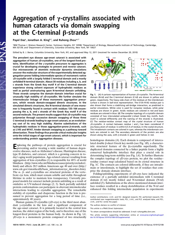
protein conformations can participate in aberrant intermolecular interactions leading to crystallin aggregation. The remarkable stability of crystallins and chaperone function in the lens is pre- sumed to prevent aggregation for a long period of time (i.e., approximately 40 years). Human gamma D crystallin (γD-crys) is the third most abun- dant γ-crystallin in the lens and a significant component of the age-onset cataract. It is primarily located in the central lens nucleus, the oldest region of the body, and is therefore one of the longest-lived proteins in the human body. As shown in Fig. 1A, γD-crys is a monomeric protein composed of two structurally homologous domains (4). Each domain is composed of interca- lated double β-sheet Greek key motifs (see Fig. 1B), a character- istic structural feature of the βγ-crystallin superfamily. The duplicated domains connected by a linker peptide form a highly conserved hydrophobic interface that plays a crucial role in determining long-term stability (see Fig. 1A). To further illustrate the complex topology of γD-crys protein, we plot the residue– residue contact map calculated based on its crystal structure in

  • Fig. 1C. The contacts are colored differently, if they are intrado-

main or interdomain, to highlight the set of residues that com- prise the domain–domain interface. Folding/unfolding experiments of γD-crys have indicated the existence of a partially unfolded intermediate with C-terminal domain (C-td) mostly folded and N-terminal domain (N-td) unfolded (5). Substitutions at the above-mentioned domain inter- face residues resulted in a sharp destabilization of the N-td and enhanced this folding intermediate population in experiments

  • Fig. 1.

(A) A cartoon representation of human γD-crystallin. The N-terminal domain (N-td) and the C-terminal domain (C-td) are shown in yellow and green, respectively. The heavy side chain of the residues at the interdomain surface is shown in ball-stick representation. The E135–R142 residue pair is also shown that forms a stabilizing salt-bridge interaction, as predicted in earlier simulations. White color is used for nonpolar residues, while polar residues are shown in green. Acidic residues are colored in red and basic residues are colored in blue. (B) The complex topology of a crystallin domain consisted of two intercalated antiparallel β-sheet Greek Key motifs. Each motif is colored differently and the naming of the strands is illustrated. (C) The residue–residue contact map of the crystal structure of human γD-crystallin. A contact between residue i and j has been considered if any heavy atom of residue i is within 6.5 A of residue j in the crystal structure. The intradomain contacts are colored in cyan, whereas the interdomain con- tacts are colored in red. The secondary elements of the protein are also shown along the axes, with β-strands in green and helices in red.

Author contributions: J.A.K. and R.Z. designed research; P.D. performed research; P.D. contributed new reagents/analytic tools; P.D., J.A.K., and R.Z. analyzed data; and P.D., J.A.K., and R.Z. wrote the paper. The authors declare no conflict of interest. This article is a PNAS Direct Submission.

1To whom correspondence should be addressed. E-mail: ruhongz@us.ibm.com.

This article contains supporting information online at www.pnas.org/lookup/suppl/ doi:10.1073/pnas.1019152108/-/DCSupplemental. 10514–10519 ∣ PNAS ∣ June 28, 2011 ∣ vol. 108 ∣ no. 26 www.pnas.org/cgi/doi/10.1073/pnas.1019152108

slide-2
SLIDE 2

(5–7). The isolated N-td is able to fold on its own into the native configuration in experiments; however, the stability of the iso- lated N-td is much lower compared to that in the full monomer (8). These results suggest that (i) the C-td stabilizes the N-td in the context of full γD-crys monomer, and (ii) the C-td interface serves as a template for the N-td folding in the full protein. Characterization of cataractogenesis in the native environment has been difficult due to the physical integrity of the lens. The direct identification of the state of aggregation precursors within the lens fiber cells or the intact lens has not been achieved due to experimental complications. However, the experimentally found partially unfolded intermediate conformation of γD-crys under- goes aggregation that competes with productive refolding, as

  • bserved during equilibrium unfolding/refolding experiments

(9, 10). This in vitro off-pathway aggregation in GdmCl at pH 7 provides a unique model for studying crystallin aggregation in

  • vivo. Atomic force microscopy indicated that the aggregated state
  • f γD-crys is ordered filament-like (10). The bis-ANS binding of

γD-crys aggregate species suggested presence of exposed hydro- phobic pockets (10). More recent experiments reveal that at low pH, γD-crys polymerize into amyloid fibrils (11), similar to those

  • bserved in neurodegenerative diseases.

Taken together, the detailed mechanism of crystallin polymer- ization is still unknown due to a lack of structural information of the monomeric and oligomeric aggregation-prone species. Map- ping the initial pathways of crystallin aggregation can provide a route toward targeted searches for therapeutic agents inhibiting pathological deposition for a number of protein deposition dis- eases including cataract. Molecular dynamics simulations (12, 13)

  • f protein models at different resolutions, from simple models

(lattice and off-lattice) (14–17) to continuum solvent models (18, 19) to all-atom explicit solvent models (20, 21), have served as a powerful tool to complement existing experimental techni- ques to advance our fundamental understanding of protein fold- ing and aggregation. In this study, using extensive atomistic mole- cular dynamics simulations performed on the IBM Blue Gene/L supercomputer we have characterized unfolding of human gam- ma D crystallin followed by oligomerization, which is consistent with the current models of cataract. A partially unfolded inter- mediate conformation strikingly similar to the experimentally

  • bserved aggregation-prone folding intermediate species is de-

tected in our ≥2 μs denaturation simulations. This unfolded intermediate species has its C-td more native-like and the N-td largely unfolded. T

  • explore the aberrant protein–protein inter-

actions promoting γD-crys aggregation, we further simulated two partially unfolded monomeric conformations together. Our ≥1 μs simulated annealing molecular dynamics simulations of the modeled γD-crys dimeric system reveal successive domain swap- ping at the C-terminal β-strands as a molecular mechanism for γ-crystallin aggregation. Thus, this simulation study uncovers a molecular picture of the unfolding and polymerization reactions associated with the current models for cataract formation. Because crystallins are model proteins for understanding β-sheet folding, these molecular pictures obtained from simulated un- folding and oligomerization of human γD-crystallin can also offer important clues to solve the so-called protein folding as well as to understand the misfolding and aggregation of β-sheets implicated in protein conformational diseases. Results and Discussion

An Intermediate State Is Populated During Unfolding of γD-crys

  • Monomer. Fig. 2A summarizes the primary events of the simulated

unfolding reaction of the native monomeric γD-crys protein performed at 425 K in 8 M aqueous urea. In this figure the time evolution of the fraction of native contacts formed for the two domains, Q(N-td) and Q(C-td) are plotted against each other, as

  • btained from different unfolding trajectories with a total simu-

lation time of approximately 2 μs. A native contact is considered to be formed between residues i and j if a heavy atom of residue i is within 6.5 Å of a heavy atom of residue j in the crystal structure. For the unfolded protein, Q(N-td) and Q(C-td) are close to 0, whereas for the folded state, both Q(N-td) and Q(C-td) are ≈1. The time evolution of Q(N-td) and Q(C-td) clearly shows sequential unfolding of two domains, consistent with experimen- tal results (5–7, 10). The unfolding of N-td always preceded that

  • f C-td, as indicated by the rapid loss of Q(N-td) compared to

Q(C-td). This higher stability of the C-td agrees well with experi- mental data (22). We also show characteristic conformations populated at different stages of unfolding in Fig. 2A. The struc- tural details of these conformations suggest that the interdomain contacts are broken before denaturation of the N-td starts [at QðC-tdÞ ¼ ∼0.6, QðN-tdÞ ¼ ∼0.6, I1 configuration]. Next, a par- tially unfolded conformation (I2) with its C-td folded ðQðC-tdÞ ¼ ∼0.4Þ and N-td largely denatured ½QðN-tdÞ < 0.3 is populated, in agreement with the experimental finding of a γD-crys folding intermediate (10). The C-td finally unfolds (to U conformation) to complete γD-crys denaturation.

Exposure of Hydrophobic Patches Within the C-td upon N-td Unfolding.

Because the structure of the I2 ensemble populated in the simu- lated unfolding is highly similar to the experimentally detected folding intermediate with a more native-like C-td and an un- folded N-td that serves as the aggregating precursor in vitro, we further analyze the structure of the I2 ensemble to identify crucial structural changes. Although the C-td remains more native-like compared to the N-td in the I2 ensemble, the C-td also experi- ences partial unfolding, as indicated by an approximately 60% reduction of the Q(C-td) from the crystal structure. We also find that, upon N-td unfolding the C-td undergoes a net approximat- ley 23% increase in solvent-exposed surface area (probed using a sphere of 1.4 Å) from the crystal structure. Recent experiments also suggested that a partial unfolding of the C-td is required for aggregation, further demonstrating the structural similarity be- tween the simulated folding intermediate and the experimentally detected aggregation-prone species (23). To map this structural change of the C-td at the residue level, the root-mean-square fluctuation (RMSF) and the %SASA increase in the I2 ensemble as well as the hydrophobicity of each residue within the C-td were calculated (Fig. 2B). The RMSF per residue plot clearly indicates that only the loop regions from Greek key motif 3 of the C-td

  • Fig. 2.

(A) SimulatedunfoldingofhumanγD-crystallin at425Kin 8M aqueous

  • urea. The fraction of native contacts formed, Q(?), for the two domains are

plotted against each other, as obtained from an aggregate of approximately 2 μs of unfolding simulations. Each point on this plot is colored from blue to red according to its time sequence during unfolding. Typical conformations populated at different stages of unfolding are also shown. (B) Structural changes of the C-td in the I2 ensemble. The RMSF in Å from the native structure, the percentageofsolvent-exposedsurfacearea(%SASA)change, and theKyte– Doolittle hydrophobicity are plotted for each residue of the C-td within the I2 ensemble consisting >1;000 conformations with QC-td ≥ 0.4 and QN-td ≤ 0.3. The regions that undergo strong conformational fluctuation and/or solvent exposure, as well as contain hydrophobic residues, are highlighted. Das et al. PNAS ∣ June 28, 2011 ∣

  • vol. 108

  • no. 26

∣ 10515

BIOPHYSICS AND COMPUTATIONAL BIOLOGY

CHEMISTRY

slide-3
SLIDE 3

become more flexible upon N-td unfolding. On the other hand, residues 132–164 from the Greek Key motif 4 that include a, b, and c strands suffer large structural fluctuation (>3 Å). Those re- sidues encompassing the interdomain interface also experience strongest solvent exposure within the C-td with a net contribution

  • f >50% to the total increase in SASA. The hydrophobicity ana-

lysis reveals presence of eight hydrophobic residues in the 132–164 region, as delineated by the widely used Kyte–Doolittle hydropho- bicity scale, in which regions with values above 0 are defined as

  • hydrophobic. T

aken together, these analyses suggest that the a, b, and c strands from the Greek key motif 4 that contain a large hydrophobic region experience large structural perturbation as well as strong solvent exposure upon unfolding of the N-td.

Simulations of Oligomerization of Aggregation-Prone Monomeric

  • Species. Because of its high structural resemblance with the ex-

perimentally detected aggregation-prone folding intermediates, the partially unfolded intermediate with its C-td more native-like and N-td largely unstructured detected in our unfolding simula- tions provides us an excellent candidate to study the mechanism

  • f intermolecular association during the competing off-pathway

polymerization reaction in vitro. To characterize the intermole- cular interactions between those aggregation-prone species of γD-crys we designed an in silico experiment. In this experiment, the conformational dynamics of one partially unfolded monomer in water is studied in presence of a second partially unfolded molecule by using extensive atomistic molecular dynamics simu- lations combined with a thermal annealing protocol (see System and Methods for details). The initial monomeric conformations for this experiment are selected from the I2 ensemble (see

  • Fig. 1C). The simulations of the dimeric γD-crys are then per-

formed in three different steps: (i) The N-tds of the monomers are first fully unfolded by thermal denaturation; (ii) the confor- mational space of the denatured N-tds is then explored by simu- lated annealing molecular dynamics; (iii) the whole system is allowed to relax at 350 K. During step i and step ii, the C-tds of the monomers remain constrained and only N-tds are allowed to

  • move. Nine different sets of dimeric systems were simulated start-

ing from different starting monomeric conformations represent- ing the I2 ensemble. In the dimeric system, the distance between the C-tds was varied from 30 Å to 56 Å. In addition, the relative

  • rientation of the monomers was also varied by changing the

monomer–monomer angle from 70° to 160°. The initial (at the end of stage i) and final conformations (at the end of stage ii and iii) for these trajectories are shown in Fig. S1. For compar- ison, the individual monomers alone were also simulated using the same protocol stated above. During this in silico experiment with modeled monomers and dimers, we consider the N-td to be unfolded, if radius of gyration, Rg, is >20 Å, and solvent acces- sible surface area (SASA) is >10;000 Å2. A collapsed state of the N-td is considered if Rg is ≤15 Å and SASA is <7;500 Å2 (the native N-td has a Rg of 11.7 Å and a SASA of 4;850 Å2). Thus, a >5 Å lowering in Rg and a >2;500 Å2 decrease in SASA define the collapse of the N-td in this study. We should also emphasize that the simulated annealing molecular dynamics simulations performed in this study are used to solely investigate the nonspe- cific collapse, but not the folding, of the N-td polypeptide chain; as such, study is impossible with current computational resources.

Collapsed N-td Forming Interdomain Contacts in the Dimeric Struc-

  • ture. The time evolution of the radius of gyration of the N-td

(see Fig. S2A) was monitored during the in silico experiment to follow the conformational change of the monomers. We find that the denatured N-tds of the partially folded monomers, both in isolation and in presence of a second monomer, undergo a col- lapse at 350 K, as indicated by the sharp decrease in both radius

  • f gyration and solvent accessible surface area. The time scale of

this simulated collapse (the first time N-td experiences a nonspe- cific collapse starting from the completely denatured state generated at the end of step i) of the N-td is approximately 20–100 ns. Fig, S2A, Top, illustrates the characteristic behavior

  • f the radius of gyration of the N-td during one such simulation
  • f the dimeric system, suggesting formation of a collapsed state

(Rg < 15 Å) from an extended state (Rg > 25 Å) of the N-td. Table S1 summarizes the initial intermonomer orientation and distance as well as the radius of gyration of the N-td in the final dimeric ensemble for all nine runs performed in this study. We found that the collapse of the N-td is favored in presence of a second monomer, if the two monomers are oriented in an anti- parallel manner (the angle between two monomers approaching 180°) and the distance between two monomers is lowered (see Table S1). This observation suggests possible formation of inter- molecular interactions facilitating the N-td collapse. Next, the number of contacts between the N-td and the C-td, NQN-C, was estimated as a function of simulation time (Fig. S2A, Bottom). An interdomain contact between the N-td and the C-td is considered, if the Cα atom of any residue i from the N-td is within 10 Å of the Cα atom of any residue from the C-td. An increase in the total number of interdomain contacts, NQN-C, was

  • bserved during the N-td collapse for the monomers as well as

for the dimers, as shown in Fig. S2A. Such an increase in NQN-C advocates the essential interaction of the N-td with certain regions of the C-td during its collapse. To investigate the time order of the collapse of the N-td and its interaction with the C-td, we have estimated the cross-correlation function between Rg and NQN-C time series. Fig. S2B shows the typical cross-correlation found between Rg of the N-td and NQN-C as a function of time lags for one I2 conformation during simulations of the isolated molecule and in presence of a second

  • conformation. The cross-correlation reaches its peak at time lag

approximately 0 for all simulations, suggesting that the collapse of the N-td is consistently accompanied by formation of contacts with the C-td for simulations of both monomers and dimers.

Mechanism for γD-protein Oligomerization. The final collapsed

conformations at the end of simulations were always found to form interdomain interactions. Those interactions were both intramonomer and intermonomer for the dimeric system—i.e., the N-td of a monomer can interact with its own C-td or with the C-td of the neighboring monomer, which is strongly determined by the initial conformations. For example, if the two monomers are aligned to each other in an antiparallel manner and they are closer to each other, more intermonomer interactions are formed between the N-td and the C-td (see Table S1). A few representative conformations from the ensemble of dimeric γD-crys with col- lapsed N-tds, which is populated at the end of our simulations, are shown in Fig. 3A (see also Fig. S1). Such conformations repre- sent stable endpoints of the simulation and are stable for tens of

  • nanoseconds. In the first example, we show a final conformation in

which both N-tds interact with the C-tds in intra- as well as in in- termolecular fashion, forming a structure similar to a close-ended

  • dimer. In the second example, one N-td forms contacts with the

C-td from the same monomer, whereas the other N-td interacts with the C-d in both intra- and intermolecular fashion. We also find conformations in which the N-td of one monomer remains denatured, whereas the N-td of the second monomer undergoes collapse by forming interchain contacts with the C-td, thus forming conformation similar to a N-terminal open-ended dimer (example 3 and 4 in Fig. 3A). These simulations thus reveal that the oligo- merization of the partially unfolded intermediates can occur through interactions at the domain interface. The final dimeric ensemble obtained from those trajectories contains structurally different domain-swapped conformations, which are either close- ended or open-ended. However, estimating the relative abun- dance and stability of those dimeric species require simulations, which is beyond the limit of the current study. From the current

10516 ∣ www.pnas.org/cgi/doi/10.1073/pnas.1019152108 Das et al.

slide-4
SLIDE 4

simulations, it appears that the close-ended domain-swapped dimer formation is favorable, when the two monomers are or- iented in an almost antiparallel manner. As the relative orienta- tion starts deviating from antiparallel, formation of more open- ended domain-swapped dimers is noticed. To examine the propensity of forming inter- or intramolecular contacts within the C-td, we analyze the average number of inter- and intramolecular contact formation per residue of the C-td in an ensemble of >1;000 final dimeric conformations that are col- lected from trajectories of runs 1–4 (Table S1) showing domain swapping, in which N-tds are collapsed (Fig. 3B). For comparison, we also plot the propensity of interdomain contact formation in the final conformational ensemble of isolated monomers. The regions from the C-td that were identified with high number of contact formation with the collapsed N-td, both in dimers and monomers, resemble the native interdomain interface. One ex- ception is the participation of the 110–120 loop in the interaction with the N-td in the isolated monomers, which becomes partially unstructured in the I2 ensemble (see Fig. 3B). In the dimeric structures, the C-td residues are found to interact with the N-td in both intra- and intermonomer fashion. Particularly, residues 135–164 comprising a, b, and c strands from the Greek Key motif 4 are frequently found to interact with the N-td in an intermole- cular manner (see Fig. 3B). Remarkably, the same region con- taining six hydrophobic residues experience partial unfolding and strong solvent exposure in the monomeric partially unfolded folding intermediate species. Thus, these findings suggest that, about 30 amino acids including three antiparallel β-strands from the C-td, which also comprise the native domain-domain inter- face, are swapped between adjacent monomers to facilitate the intermolecular association of partially unfolded aggregation- prone conformations of γD-crys. As a result, a “native-like” in- terdomain surface is formed in the dimers. In the current context, presence of a native-like interdomain surface in the dimeric sys- tem suggests participation of residues from the C-td, which also comprise the native interdomain surface (see Fig. 1C), in the for- mation of intermonomer linkage. As seen in the crystal structure, the interdomain contacts primarily involve a, b, c, and d strands of the Greek Key motif 4 (Fig. 1C). From the simulated dimric en- semble, it is clear that a, b, and c strands of the Greek Key motif 4 significantly contribute to the formation of the intermonomer surface, thus resulting into a native-like interdomain surface in the dimers (Fig. 3B). As mentioned earlier, six of those residues are strongly hydro-

  • phobic. T
  • test if those hydrophobic residues indeed play a key

role in promoting dimerization, we have substituted in silico two of those six residues, L145 and M147, with alanine and simu- lated the dimerization of the double mutant protein using similar protocol as used for the wild-type protein. Strikingly, the final ensemble for the double mutant shows less inter- as well as in- trachain interdomain contact formation compared to the wild- type protein. For direct comparison, simulations of the double mutant and wild-type dimers were started from identical initial conformations, except that the residues L145 amd M147 were substituted with alanines. Fig. 3C shows the residues from the Greek Key motif 4 of the C-td of one monomer that form contact with the N-td of the second molecule during the last 25 ns of one representative MD trajectory, for the wild-type (left) and for the double mutant (right). Clearly, the intermolecular swap of the ab hairpin from the Greek Key motif 4 takes place to a much lower extent in the double mutant compared to the wild-type protein. The residues L145 and M147 are major components of the hydrophobic interdomain interface in the monomeric γD-crys, which significantly contributes to the overall native stability. Our simulations suggest that substituting those two residues with less hydrophobic ones may hinder domain swapping as a pathway toward dimerization in γD-crystallin.

Role of Interdomain Surface in Crystallin Stability. All known βγ-

crystallins from extant vertebrate species have duplicated Greek key domains that are presumably evolved by gene duplication and gene fusion from an ancestral single domain crystallin (24, 25), as suggested by the high sequence and structural similarity of the two domains. In fact, such single domain crystallins have been identified—for example, in the Sea squirt Ciona (26). These

  • rganisms may represent descendants of lineages that are candi-

dates for the origin of the vertebrates. It is also noteworthy that most βγ-crtstallins exhibit differential domain stability (27), sug- gesting that addition of a second domain and the interdomain interface to the ancestral single domain protein contribute to the overall stability of the full-length protein. In fact, stability comparisons of the isolated domains with the full γD-crys mono- mer indicated that the domain interface contributes a ΔGH2O of approximately 4.2 kcal∕mol to the stability of the full monomer (22). This additional stability conferred by the interfacial interac- tions between the duplicated domains is likely very important for maintaining the long life time of proteins of the lens nucleus, and thus explains the selection for duplicated forms. The crucial role of the interdomain interface in the folding, stability, and aggregation of βγ-crystallins is supported by experi- ments (5–7, 28). In a previous unfolding simulation study of the isolated domains of γD-crys, we have shown that the a and b

  • Fig. 3.

Domain-swapped dimersof γD-crys.(A) Final domain- swapped dimer configurations are shown from three differ- ent runs. The folded C-td and the unfolded N-td of one mono- mer are colored in green and yellow, respectively. For the second monomer, the folded C-td and the unfolded N-td are colored in blue and red, respectively. (B) The average number of contacts with the collapsed N-td is plotted for each residue within the C-td, as obtained from an ensemble of >1;000 conformations, for both monomeric and dimeric sys- tems.Thoseconformationsarepopulatedduringthelast 50ns

  • f the simulations at approximately 350 K, in which the N-tds

are collapsed. The results obtained from isolated monomer simulations are colored in black. The probabilities of intra-, inter-, and total (intra- þ inter-) interdomain contact forma- tion per residue in dimers are plotted in cyan, red, and gray,

  • respectively. (C) The number of interchain contacts formed

between the N-td and every residue of the Greek key motif 4 during the last 25 ns of MD at 350 K, for (left) the wild-type dimer and for (right) the double mutant dimer. The number

  • f interchain interdomain contacts is colored according to

the color scale shown. The alanine substitution sites are indi- cated with an red arrow, where a decrease in intermolecular interaction is noticed in the double mutant system. Results for

  • ne representative trajectory are shown.

Das et al. PNAS ∣ June 28, 2011 ∣

  • vol. 108

  • no. 26

∣ 10517

BIOPHYSICS AND COMPUTATIONAL BIOLOGY

CHEMISTRY

slide-5
SLIDE 5

strands from the Greek Key motif 4 comprising the interdomain interface are the most stable structure within full γD-crys (29). We also uncovered that a Glu-Arg salt-bridge at the topologically equivalent positions of residues E135 and R142 (see Fig. 1A and

  • ref. 29) plays a significant role in determining the stability of a

Greek Key motif. Disrupting the E135-R142 salt-bridge in silico resulted in destabilizing the interdomain interface and facili- tated the N-td unfolding (29). Similarly, an Arg14 to Cys (R14C) mutation on γD-crys breaking the E7-R14 salt-bridge in the Greek Key motif 1, which is associated with a juvenile-onset hereditary cataract (30), triggers formation of intermolecular aggregates in physiological pH.

Domain Swapping as a Molecular Mechanism for Crystallin Polymer-

  • ization. Domain swapping (31, 32) has been recognized as an

aggregation mechanism for a number of proteins—for example, human prion protein (33) and β2-microglobulin (34). Aggrega- tion by domain swapping in an open-ended fashion has been

  • bserved for cystatin C (35). Successive domain swapping of a

single domain has been previously suggested as a mechanism for polymerization on the basis of the dimeric and/or trimeric structures of Serpin (36), RNAse A (37), and cytochrome C (38). Domain swapping of an α-helix has been reported in the minor dimeric structure of RNase A (39) and in staphylococcal nuclease (40), as well as in the dimeric and trimeric structures of cyto- chrome c (38). The major dimeric component of RNase A is formed by swapping of the C-terminal β-strands (41). The crystal structure of a stable dimer of serpin, a protein family that forms large stable multimers leading to intracellular accretion and dis- ease, revealed a domain-swapped structure of two long antipar- allel β-strands (36). Domain-swapped trimeric structures have been also reported for Barnase (42) and an antibody fragment (43), but the mechanism of polymerization was not elucidated. Domain-swapped polymers have been detected for a number

  • f functional proteins as well, such as T7 helicase, RecA, and car-

bonic anhydrase (32). Solved structures of the βB2-crystallins show natural close-ended domain-swapped dimer, in which the N-td interacts with the C-td intermolecularly (44). Based on the presence of duplicated domains and the crucial role of native interdomain surface in γD-protein stability, together with the in vitro off-pathway aggregation reaction of the folding intermedi- ate, domain swapping has been proposed as a plausible model for γD-crys polymerization. However, aggregation by a domain swap mechanism has not been experimentally observed for the

  • crystallins. Our large-scale simulations provide direct evidence

supporting this model, in which the aggregation-prone folding in- termediate is found to form domain-swapped dimeric structures. Structures similar to both open-ended and close-ended domain- swapped dimers were detected in our simulations, which suggests successive domain swapping as a possible mechanism for crystallin aggregation in aged lens leading to cataract (Fig. 4). We identify structural components of γD-crystallin that are prone to domain

  • swapping. This information can help us to design drug-like mole-

cules that can prevent cataractogenesis in eyes. For example, small molecules that can attach to the native domain–domain interface and, therefore, hinder unfolding of the N-td can serve as drugs to avoid cataract. Those molecules can provide additional stability to the interdomain surface and prevent the C-terminal β-strands from solvent exposure. In addition, based on simulations we propose two potential mutation sites, L145 and M147, in which alanine substitution can retard dimerization via domain swapping. The findings of this simulation study, together with the pre- vious experiments, shed important insights onto the pathways

  • f crystallin aggregation, which can be connected to the more

general theory of protein aggregation, suggesting the critical role

  • f an aggregation-prone monomeric species (termed as I2 in this

study, often referred as N* in literature; see refs. 45 and 46 and references therein). The role of N* state has been implicated in the aggregation pathways of transthyretin, prion proteins, and a variety of amyloid proteins including aβ protein. Any changes in the solution or environmental conditions (e.g., heat, UV rays) or mutations that destabilize the native γ-crystallin will trigger the formation of the aggregation-prone partially unfolded mono- meric folding intermediates (N*). In such circumstances, succes- sive domain swapping of the C-terminal strands may lead to polymerization in a highly crowded lens environment (Fig. 4). The emerging role of the native interdomain surface in triggering polymerization indicate that the crystallin aggregation in aged eye lens is an evolutionary cost of the long-term native state stability, which, in turn, is determined by its complex domain architecture. Because the successive intermolecular protein association by means of β-sheet expansion (47) is also the mechanism underlying a multitude of diseases including Alzheimer’s disease, Hunting- ton disease, Parkinson disease, and the prion encephalopathies, the detailed picture of crystallin unfolding followed by polymer- ization revealed in this study may provide valuable information toward understanding those conformational diseases (48). Conclusion Large-scale atomistic simulations were used to reveal the mole- cular structures of the aggregation-prone intermediate species of human γD-crystallin, an eye lens protein implicated in catarac-

  • teogenesis. The partially unfolded conformation with a mostly

folded C-td and a largely denatured N-td identified in the unfold- ing simulations has a structure strikingly similar to the aggrega- tion-prone folding intermediate species observed in experiments. A detailed structural characterization of this pathogenic mono- meric state N* reveals the presence of a solvent-exposed partially unfolded region within the more native-like C-td, which is ap- proximately 30 amino acids long and contains eight hydrophobic

  • residues. This region includes the first three antiparallel strands

from the Greek Key motif 4, which also encompasses the inter- domain interface crucial for native γD-crystallin stability. We further simulated the intermolecular association of these mono- meric partially unfolded aggregation precursors (N*) to elucidate the mechanism of γD-crystallin aggregation. The denatured N-tds experience a distinct nonspecific collapse that is essentially accompanied by formation of specific interactions with the C-tds. As a result, domain-swapped dimeric conformations were popu- lated in our simulations. The partially unfolded and solvent- exposed region encompassing a, b, and c strands from the Greek Key motif 4 in N* ensemble was found to be exchanged between adjacent monomers, forming a native-like interdomain interface

Open-ended domain- swapped dimer Unfolding

  • f N-td

Close-ended domain- swapped dimer I2 (N*)

A C B D E

Polymerization via domain swapping

  • Fig. 4.

Schematic summary of human γD-crys polymerization. (A) Crystal structure of human γD-crys. (B) Simulated monomeric aggregation precursor (I2), often referred as N* in the general mechanism of protein aggregation in

  • literature. (C) Simulated structure of open-ended domain-swapped dimer.

(D) Simulated structure of close-ended domain-swapped dimer. (E) Model

  • f human γD-crys hexamer formed via domain swapping. The coloring

scheme for the dimer system is same as in Fig. 3. 10518 ∣ www.pnas.org/cgi/doi/10.1073/pnas.1019152108 Das et al.

slide-6
SLIDE 6

in the dimeric structures. We also observed that alanine substitu- tions of the hydrophobic residues in those aggregation-prone β-strands, such as L145 and M147, located at the end of the ab hairpin of the Greek Key motif 4 hinders domain swapping as a pathway toward dimerization. In an older lens, the native pro- teins may aggregate by propagating domain swapping in an open- ended fashion along multiple partially unfolded monomers leading to cataract. This simulation study thus provides critical insights onto the molecular mechanism of initial stages of age-

  • nset cataract formation, which is also important toward under-

standing other protein aggregation diseases. System and Methods The initial structure of the wild-type γD-crystallin protein (see

  • Fig. 1A) containing 173 residues has been taken from the crystal

structure deposited in the Protein Data Bank (PDB ID code 1HK0). The molecular system for unfolding studies was prepared by immersing the full monomer in 8M urea. The system contained approximately 7,650 water molecules and approximately 1,775 urea molecules—a total of approximately 40,000 atoms. The par- tially unfolded intermediate conformations were generated by unfolding the full γD-crys monomer at 425 K using 8 M aqueous urea as a denaturant. Typical length of such unfolding simulations was >300 ns. Details of the simulation setup and analysis of γD-crys unfolding can be also found in SI Text and ref. 29. T

  • explore the oligomerization pathway(s) of γD-crys, two par-

tially unfolded γD-crys molecules were placed together in a box

  • f size ∼100 Å × 100 Å × 100 Å containing approximately 26,000

TIP3P water molecules. Simulations were performed with differ- ent relative orientation ranging from 70° to 160° and with distance between two C-tds ranging from 30 Å to 56 Å. The system con- taining a total of approximately 100,000 atoms then went through a sophisticated annealing process (details in SI Text). Finally, we allowed the complete system to relax at 350 K for >50 ns. This in silico experiment on simulating dimeric structures was repeated nine times starting from different configurations, in which the monomer conformations as well as the intermonomeric distance and orientation were varied. The partially folded monomers alone were also simulated in water using the same protocol to compare the intramolecular interactions with the intermolecular interactions present in the dimeric system. L144 and M146 were mutated in silico to alanine to create the initial structure of the double mutant dimer, which was then simulated using a similar protocol stated above. The aggregate MD simulation time for both wild-type and the mutant oligomers is approximately 2 μs.

  • 1. Benedek GB (1997) Cataract as a protein condensation disease: The Proctor lecture.

Invest Ophthalmol Vis Sci 38:1911–1921.

  • 2. Aarts HJM, Lubsen NH, Schoenmakers JGG (1989) Crystallin gene expression during rat

lens development. Eur J Biochem 183:31–36.

  • 3. Lampi KJ, Shih M, Ueda Y, Shearer TR, David LL (2002) Lens proteomics: Analysis of rat

crystallin sequences and two-dimensional electrophoresis map. Invest Ophthalmol Vis Sci 43:216–224.

  • 4. Basak A, et al. (2003) High-resolution X-ray crystal structures of human gamma

D crystallin (1.25 angstrom) and the R58H mutant (1.15 angstrom) associated with aculeiform cataract. J Mol Biol 328:1137–1147.

  • 5. Flaugh SL, Kosinski-Collins MS, King J (2005) Interdomain side-chain interactions in

human gamma D crystallin influencing folding and stability. Protein Sci 14:2030–2043.

  • 6. Flaugh SL, Kosinski-Collins MS, King J (2005) Contributions of hydrophobic domain

interface interactions to the folding and stability of human gamma D-crystallin. Protein Sci 14:569–581.

  • 7. Flaugh SL, Mills IA, King J (2006) Glutamine deamidation destabilizes human gamma

D-crystallin and lowers the kinetic barrier to unfolding. J Biol Chem 281:30782–30793.

  • 8. Iliopoulos I, et al. (2003) Evaluation of annotation strategies using an entire genome
  • sequence. Bioinformatics 19:717–726.
  • 9. Kosinski-Collins MS, Flaugh SL, King J (2004) Probing folding and fluorescence quench-

ing in human gamma D crystallin Greek key domains using triple tryptophan mutant

  • proteins. Protein Sci 13:2223–2235.
  • 10. Kosinski-Collins MS, King J (2003) In vitro unfolding, refolding, and polymerization of

humangammaDcrystallin,aproteininvolvedincataractformation.ProteinSci12:480–490.

  • 11. Papanikolopoulou K, et al. (2008) Formation of amyloid fibrils in vitro by human

gamma D-crystallin and its isolated domains. Mol Vis 14:81–89.

  • 12. Warshel A (2002) Molecular dynamics simulations of biological reactions. Acc Chem

Res 35:385–395.

  • 13. Karplus M, McCammon JA (2002) Molecular dynamics simulations of biomolecules. Nat

Struct Mol Biol 9:646–652.

  • 14. Wolynes PG, Onuchic JN, Thirumalai D (1995) Navigating the folding routes. Science

267:1619–1620.

  • 15. Dill KA, et al. (1995) Principles of protein folding—a perspective from simple exact
  • models. Protein Sci 4:561–602.
  • 16. Das P, et al. (2005) Characterization of the folding landscape of monomeric lactose

repressor: Quantitative comparison of theory and experiment. Proc Natl Acad Sci USA 102:14569–14574.

  • 17. Das P, Matysiak S, Clementi C (2005) Balancing energy and entropy: A minimalist

model for the characterization of protein folding landscapes. Proc Natl Acad Sci USA 102:10141–10146.

  • 18. Feig M, Brooks CL (2004) Recent advances in the development and application of

implicit solvent models in biomolecule simulations. Curr Opin Struct Biol 14:217–224.

  • 19. Zhou R, Berne BJ (2002) Can a continuum solvent model reproduce the free energy

landscape of a beta -hairpin folding in water? Proc Natl Acad Sci USA 99:12777–12782.

  • 20. Simmerling C, Strockbine B, Roitberg AE (2002) All-atom structure prediction and

folding simulations of a stable protein. J Am Chem Soc 124:11258–11259.

  • 21. Snow CD, Nguyen H, Pande VS, Gruebele M (2002) Absolute comparison of simulated

and experimental protein-folding dynamics. Nature 420:102–106.

  • 22. Mills IA, Flaugh SL, Kosinski-Collins MS, King JA (2007) Folding and stability of the

isolated Greek key domains of the long-lived human lens proteins gammaD-crystallin and gammaS-crystallin. Protein Sci 16:2427–2444.

  • 23. Moreau KL, King J (2009) Hydrophobic core mutations associated with cataract devel-
  • pment in mice destabilize human gamma D-crystallin. J Biol Chem 284:33285–33295.
  • 24. Lubsen NH, Aarts HJM, Schoenmakers JGG (1988) The evolution of lenticular proteins- the

beta-crystallin and gamma-crystallin super gene family. Prog Biophys Mol Biol 51:47–76.

  • 25. Piatigorsky J (2003) Crystallin genes: Specialization by changes in gene regulation may

precede gene duplication. J Struct Funct Genomics 3:131–137.

  • 26. Shimeld SM, et al. (2005) Urochordate beta gamma-crystallin and the evolutionary
  • rigin of the vertebrate eye lens. Curr Biol 15:1684–1689.
  • 27. Mayr EM, Jaenicke R, Glockshuber R (1997) The domains in gamma B-crystallin:

Identical fold-different stabilities. J Mol Biol 269:260–269.

  • 28. Palme S, Slingsby C, Jaenicke R (1997) Mutational analysis of hydrophobic domain

interactions in gamma B-crystallin from bovine eye lens. Protein Sci 6:1529–1536.

  • 29. Das P, King JA, Zhou R (2010) β-strand interactions at the domain interface critical for

the stability of human lens γD-crystallin. Protein Sci 19:131–140.

  • 30. Pande A, Gillot D, Pande J (2009) The cataract-associated R14C mutant of human

gamma D-crystallin shows a variety of intermolecular disulfide cross-links: A Raman spectroscopic study. Biochemistry 48:4937–4945.

  • 31. Melanie J, Bennett MPSDE (1995) 3D domain swapping: A mechanism for oligomer
  • assembly. Protein Sci 4:2455–2468.
  • 32. Liu Y, Eisenberg D (2002) 3D domain swapping: As domains continue to swap. Protein

Sci 11:1285–1299.

  • 33. Knaus KJ, et al. (2001) Crystal structure of the human prion protein reveals a

mechanism for oligomerization. Nat Struct Biol 8:770–774.

  • 34. Eakin CM, Attenello FJ, Morgan CJ, Miranker AD (2004) Oligomeric assembly of

native-like precursors precedes amyloid formation by beta-2 microglobulin. Biochemistry 43:7808–7815.

  • 35. Wahlbom M, et al. (2007) Fibrillogenic oligomers of human cystatin C are formed by

propagated domain swapping. J Biol Chem 282:18318–18326.

  • 36. Yamasaki M, Li W, Johnson DJD, Huntington JA (2008) Crystal structure of a stable

dimer reveals the molecular basis of serpin polymerization. Nature 455:1255–1258.

  • 37. Sambashivan S, Liu YS, Sawaya MR, Gingery M, Eisenberg D (2005) Amyloid-like fibrils
  • f ribonuclease A with three-dimensional domain-swapped and native-like structure.

Nature 437:266–269.

  • 38. Hirota S, et al. (2010) Cytochrome c polymerization by successive domain swapping at

the C-terminal helix. Proc Natl Acad Sci USA 107:12854–12859.

  • 39. Liu Y, Hart PJ, Schlunegger MP, Eisenberg D (1998) The crystal structure of a 3D

domain-swapped dimer of RNase A at a 2.1-Å resolution. Proc Natl Acad Sci USA 95:3437–3442.

  • 40. Green SM, Gittis AG, Meeker AK, Lattman EE (1995) One-step evolution of a dimer

from a monomeric protein. Nat Struct Mol Biol 2:746–751.

  • 41. Liu YS, Gotte G, Libonati M, Eisenberg D (2002) Structures of the two 3D

domain-swapped RNase A trimers. Protein Sci 11:371–380.

  • 42. Zegers I, Deswarte J, Wyns L (1999) Trimeric domain-swapped barnase. Proc Natl Acad

Sci USA 96:818–822.

  • 43. Pei XY, Holliger P, Murzin AG, Williams RL (1997) The 2.0-A resolution crystal structure
  • f a trimeric antibody fragment with noncognate VH-VL domain pairs shows a

rearrangement of VH CDR3. Proc Natl Acad Sci USA 94:9637–9642.

  • 44. Smith MA, Bateman OA, Jaenicke R, Slingsby C (2007) Mutation of interfaces in

domain-swapped human beta B2-crystallin. Protein Sci 16:615–625.

  • 45. Straub JE, Thirumalai D Toward a molecular theory of early and late events in

monomer to amyloid fibril formation. Annu Rev Phys Chem 62:437–463.

  • 46. Straub JE, Thirumalai D Principles governing oligomer formation in amyloidogenic
  • peptides. Curr Opin Struct Biol 20:187–195.
  • 47. Harrison RS, Sharpe PC, Singh Y, Fairlie DP (2007) Reviews of Physiology, Biochemistry

and Pharmacology, eds K Kramer, O Krayer, E Lehnartz, Av Muralt, and HH Weber (Springer, Berlin), 159, pp 1–77.

  • 48. Carrell RW, Lomas DA (1997) Conformational disease. Lancet 350:134–138.

Das et al. PNAS ∣ June 28, 2011 ∣

  • vol. 108

  • no. 26

∣ 10519

BIOPHYSICS AND COMPUTATIONAL BIOLOGY

CHEMISTRY

slide-7
SLIDE 7

Supporting Information

Das et al. 10.1073/pnas.1019152108

SI Text

Method Details. To study dimerization, the system containing a

total of approximately 100;000 atoms was gradually heated to 1,000 K followed by a ∼10 ns equilibration to completely dena- ture the N-td (Rg > 20 Å). Next, we adopted a simulated anneal- ing protocol to sample the conformational space of the denatured N-tds. In this protocol, during approximately 65 ns MD, the tem- perature of the system was lowered from 1,000 K to 500 K using a step size of 25 K and simulation length/step equal to 2 ns. The system is further cooled down to 350 K with a 25 K step size and a simulation length/step of 5 ns. During the heating and the cooling processes, the C-td backbone remained fixed. All molecular dynamics simulations were performed using NAMD2 molecular modeling package (S1) and CHARMM22 (parameter set c32b1) force field (S2) with a 2 fs time step. The particle-mesh Ewald (PME) method was used for the long-range electrostatic interactions, while the van der Waals interactions were treated with a cutoff distance of 12 Å. The un- folding simulations were performed in an NPTensemble at 425 K and 1 atm, whereas all simulations of partially unfolded mono- mers/dimers were performed in NVT ensemble.

  • 1. Kumar S, et al. (2008) Scalable molecular dynamics with NAMD on Blue Gene/L. IBM J

Res Dev 52:177–188.

  • 2. MacKerell AD, et al. (1998) All-atom empirical potential for molecular modeling and

dynamics studies of proteins. J Phys Chem B 102:3586–3616.

  • Fig. S1.

The initial (at the end of stage i) and final (at the end of stage iii) conformations for six different modeled wild-type dimer systems are shown. The initial interchain orientations and distances for those dimer systems are indicated. The folded C-td and the unfolded N-td of one monomer are colored in green and yellow, respectively. For the second monomer, the folded C-td and the unfolded N-td are colored in blue and red, respectively. Das et al. www.pnas.org/cgi/doi/10.1073/pnas.1019152108 1 of 2

slide-8
SLIDE 8
  • Fig. S2.

Dimerization of the partially unfolded monomers. (A) The radius of gyration, Rg, (in Å) and the number of interdomain contacts, NQN-C, are plotted as a function of simulation time for two different monomers during steps ii and iii in a dimerization study. The results for monomers are colored differently. A plateau of Rg at approximately 15 Å indicates the collapse of the initially unfolded N-td. (B) The typical cross-correlation found between Rg of the N-td and NQN-C as a function of time lags for one particular I2 conformation during simulations of the isolated molecule and in presence of a second conformation.

Table S1. Summary of the initial and final states of all nine runs Run θM1-M2 dM1-M2 (Å) End state of M1 End state of M2 Conclusion Rg(N-td) (Å) Interdomain contact Swapping

  • f C-td

Rg(N-td) (Å) Interdomain contact Swapping

  • f C-td

1 153 40 14.5 Yes Yes 13.5 No Yes Close-ended domain-swapping 2 134 50 13.9 Yes Yes 14.5 No No Open-ended domain-swapping 3 128 30 15.0 Yes No 14.0 No Yes Open-ended domain-swapping 4 157 50 15.0 Yes Yes 18.6 Yes No Open-ended domain swapping 5 74 30 13.6 Yes No 14.0 Yes No No significant domain swapping 6 71 37 26.0 Yes No 19.0 Yes No No domain swapping 7 150 56 17.0 Yes No 18.0 Yes No No domain swapping 8 151 47 14.5 No Yes 15.0 Yes Yes Close-ended domain swapping 9 161 51 13.0 Yes Yes 14.7 No No Open-ended domain swapping Initial intermonomer orientation and distance, as well as final N-td radius of gyration, interdomain contact formation, and C-td domain swapping. Data collected from the final dimeric ensemble for all nine runs performed in this study.

Das et al. www.pnas.org/cgi/doi/10.1073/pnas.1019152108 2 of 2