
SLIDE 1
9/29/2017 1
- E. Michael Lewiecki, MD
Director, New Mexico Clinical Research & Osteoporosis Center Director, Bone TeleHealth ECHO University of New Mexico Health Sciences Center Albuquerque, New Mexico, USA
DXA Best Practices
Dual-energy X-ray Absorptiometry: DXA
- Bone Mineral Density (BMD)
– Diagnosis – Fracture Risk (including FRAX/TBS) – Monitor
- Vertebral Fracture Assessment (VFA)
- Trabecular Bone Score (TBS)
- Hip Structural Analysis (HSA)
- Body Composition (Body Comp)
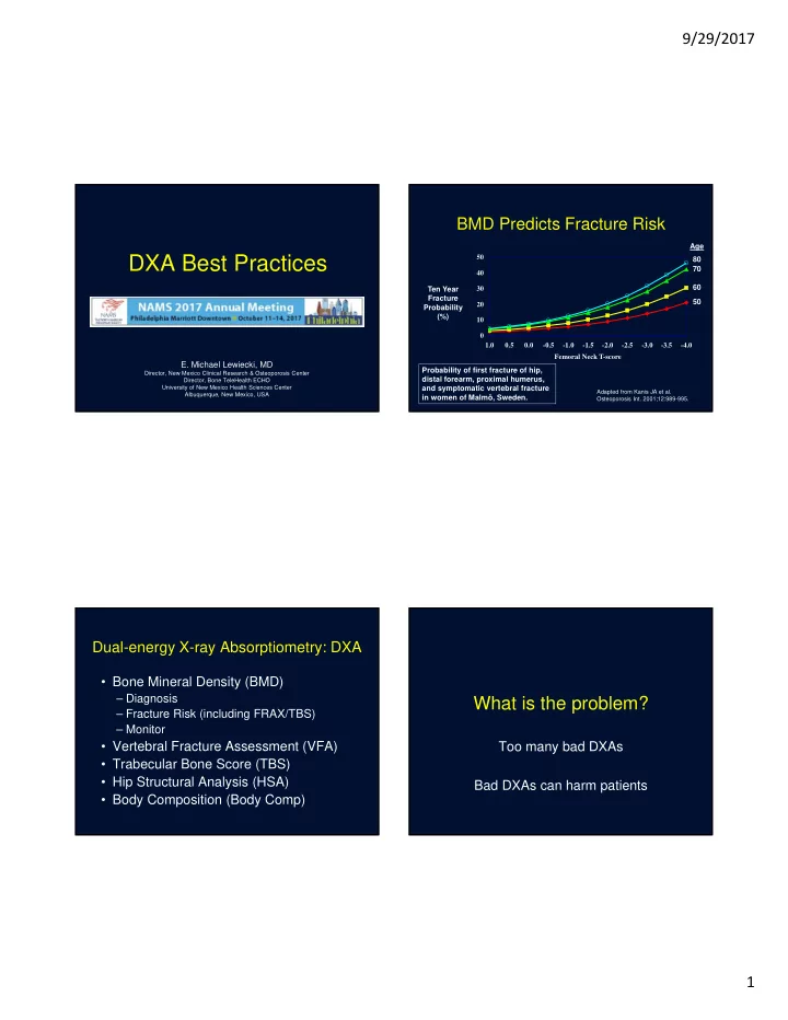
BMD Predicts Fracture Risk
Adapted from Kanis JA et al. Osteoporosis Int. 2001;12:989-995.
10 20 30 40 50 1.0 0.5 0.0
- 0.5
- 1.0
- 1.5
- 2.0
- 2.5
- 3.0
- 3.5
- 4.0
Femoral Neck T-score Ten Year Fracture Probability (%) Age 80 70 60 50 Probability of first fracture of hip, distal forearm, proximal humerus, and symptomatic vertebral fracture in women of Malmö, Sweden.