
International Journal of Oral Health and Medical Research | ISSN 2395-7387 | MAY-JUNE 2016 | VOL 3 | ISSUE 1
122 CASE REPORT
Lakhani H et al.: Unicystic Ameloblastoma in the Anterior Maxilla
Correspondence to:
- Dr. Lakhani Himanshu, Post graduate, Dept Of Oral
Medicine And Radiology Contact Us: www.ijohmr.com
An Unusual Presentation of Unicystic Ameloblastoma in the Anterior Maxilla
Himanshu Lakhani1, A.G.Annaji2, M.Manjunath3, Khalida Shaik Begum4, Anjana Ramanathan5,
- Abhinetra. M.S6
Ameloblastoma is the most common odontogenic tumor . Unicystic ameloblastoma is considered to be a variant of the solid or multicystic type. It is a less aggressive tumor with variable recurrence rate. Unicystic lesions are the ones that show clinical, radiographic and gross features of a cyst but on histologic examination shows a typical ameoblastomatous epithelium which lines a part of the cyst cavity, with or without luminal and/or mural tumor growth. We report a case of unicystic mural ameloblastoma in 21 year old male with swelling in the right upper front region of the face since 2 months. Fine needle aspiration yielded no fluid. Radiographs revealed a multilocular radiolucency in the right anterior maxillary region with buccal cortical plate expansion. KEYWORDS: Ameloblastoma, Unicystic, Mural, Maxilla
AA
aaaasasasssRobinson defined Ameloblastoma as “Unicystic, nonfunctional, intermittent in growth, anatomically benign and clinically persistent.” They are tumors of
- dontogenic epithelial origin, they may arise from basal
cells of the oral mucosa, an odontogenic cyst lining or a developing enamel organ.1 About 85% of the conventional Ameloblastomas occur in the mandible, most often in the molar-ascending ramus area whereas 15% occur in the maxilla, usually in the posterior region. The tumor is often asymptomatic and smaller lesions are usually detected only during a radiographic examination. A painless swelling or expansion of the jaw is the usual clinical presentation.1 They are usually discovered around fourth and fifth decades of life with the exception of the Unicystic variant which is more common in second and third decade of life.2 Radiographically, Ameloblastomas can either present as a unilocular or a multilocular radiolucency. The lesion is
- ften described as having a “soap-bubble” appearance
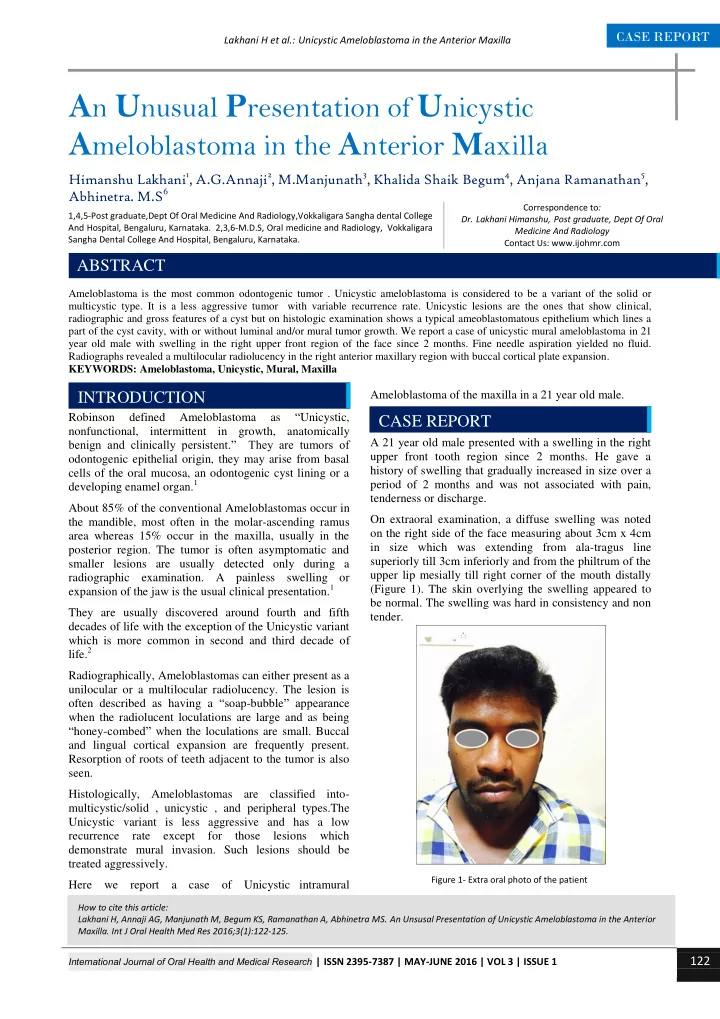
when the radiolucent loculations are large and as being “honey-combed” when the loculations are small. Buccal and lingual cortical expansion are frequently present. Resorption of roots of teeth adjacent to the tumor is also seen. Histologically, Ameloblastomas are classified into- multicystic/solid , unicystic , and peripheral types.The Unicystic variant is less aggressive and has a low recurrence rate except for those lesions which demonstrate mural invasion. Such lesions should be treated aggressively. Here we report a case of Unicystic intramural Ameloblastoma of the maxilla in a 21 year old male. A 21 year old male presented with a swelling in the right upper front tooth region since 2 months. He gave a history of swelling that gradually increased in size over a period of 2 months and was not associated with pain, tenderness or discharge. On extraoral examination, a diffuse swelling was noted
- n the right side of the face measuring about 3cm x 4cm
in size which was extending from ala-tragus line superiorly till 3cm inferiorly and from the philtrum of the upper lip mesially till right corner of the mouth distally (Figure 1). The skin overlying the swelling appeared to be normal. The swelling was hard in consistency and non tender.
How to cite this article: Lakhani H, Annaji AG, Manjunath M, Begum KS, Ramanathan A, Abhinetra MS. An Unsusal Presentation of Unicystic Ameloblastoma in the Anterior
- Maxilla. Int J Oral Health Med Res 2016;3(1):122-125.
INTRODUCTION
1,4,5-Post graduate,Dept Of Oral Medicine And Radiology,Vokkaligara Sangha dental College And Hospital, Bengaluru, Karnataka. 2,3,6-M.D.S, Oral medicine and Radiology, Vokkaligara Sangha Dental College And Hospital, Bengaluru, Karnataka.
ABSTRACT CASE REPORT
Figure 1- Extra oral photo of the patient