Disclosures Nothing to Disclose Interstitial Lung Disease: - - PowerPoint PPT Presentation

disclosures
SMART_READER_LITE
LIVE PREVIEW

Disclosures Nothing to Disclose Interstitial Lung Disease: - - PowerPoint PPT Presentation

6/22/2018 Disclosures Nothing to Disclose Interstitial Lung Disease: Decoding the Alphabet Soup Rupal J. Shah, MD MS Director, Interstitial Lung Disease Clinic University of California, San Francisco Outline What is ILD?


slide-1
SLIDE 1

6/22/2018 1

Interstitial Lung Disease: Decoding the Alphabet Soup

Rupal J. Shah, MD MS Director, Interstitial Lung Disease Clinic University of California, San Francisco

Disclosures

  • Nothing to Disclose

Outline

  • What is ILD?
  • Diagnostic Approach
  • Specific types of ILD
  • Available therapies
  • Cases
slide-2
SLIDE 2

6/22/2018 2

What is the pulmonary interstitium?

  • Anatomic space that is lined by

epithelial and endothelial cells

  • Contains collagen, elastin, reticulin,

ECM

  • Also in the connective tissue of the

lung (interlobular septa, visceral pleura, peribronchovascular sheaths)

Not Just Interstitial

  • Misnomer because many ILD’s affect the

airways, parenchyma, blood vessels and pleura

  • More accurately described as diffuse

parenchymal lung disease

  • Over 100 types of ILD’s

Farrand, E et al The hospitalized patient with interstitial lung disease: A hospitalist primer J Hosp Med 2017

Epidemiology

Lederer DJ, et al Idiopathic pulmonary fibrosis NEJM 2018

slide-3
SLIDE 3

6/22/2018 3

Diagnosis

  • Challenge: Presentation is usually nonspecific
  • Average time from symptom onset to

diagnosis: 1-2 years

  • Early recognition is important!
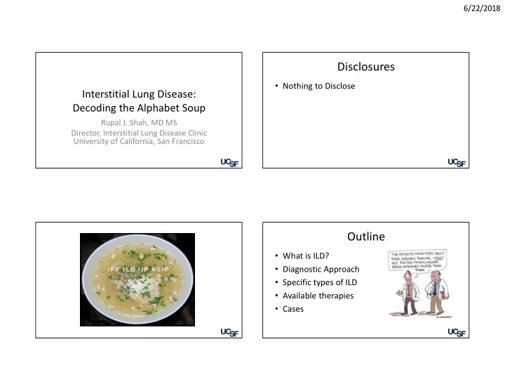
Outline

  • What is ILD?
  • Diagnostic Approach
  • Specific types of ILD
  • Available therapies
  • Cases

67 yo M with progressive dyspnea and

  • cough. Treated with abx for bronchitis.

40 pack year smoker, quit 6 years ago. On exam, late inspiratory crackles, +clubbing. CXR shows increased basilar reticulation. What additional historical information is most likely to assist in establishing a diagnosis? A. Allergy History B. Family History C. Occupational History D. Travel History

A l l e r g y H i s t

  • r

y F a m i l y H i s t

  • r

y O c c u p a t i

  • n

a l H i s t

  • r

y T r a v e l H i s t

  • r

y

0% 0% 85% 15%

Clinical Evaluation: History

12

Elements Examples Demographics

Age, IPF > 50

Time course

Acute, sub-acute, chronic

Extra-pulmonary symptoms of CTD

Raynauds, rash, inflammatory arthritis, proximal muscle weakness, dry eyes/mouth

Smoking history

DIP, RB-ILD, LCH, AEP

Medications/Radiation

Nitrofurantoin, Amiodarone, methotrexate, chemotherapy, radiation 77 medications (pneumotox.com)

HP exposures (home, work, hobbies)

Avian (birds, down), molds (water damage, swamp cooler), mycobacteria (indoor hot tub, metal working fluid)

Occupational exposures

Asbestos, beryllium, metal dusts

Family history of ILD

Early graying, cryptogenic cirrhosis, bone marrow disorders Travis, WD et al An official ATS/ERS statement: Update of the international multidisciplinary classification of IIP AJRCCM 2013

slide-4
SLIDE 4

6/22/2018 4

Clinical Evaluation: Physical Exam

13

Elements Examples Face and mucus membranes

Scleritis, saliva pool & dentition, oral ulcers, leukoplakia, mouth

  • pening, squared off telangectasias, fibrofolliculomas, malar rash

Lungs

Dry crackles (listen at bases or may miss early disease!) Inspiratory squeaks (bronchiolitis)

Cardiac

Signs of pulmonary hypertension

Hands

Clubbing, mechanics hands, sclerodactyly, nail bed capillaries, palmar telangectasias, Gottrons papules, synovitis and deformities, dystrophic nails

Neuro

Proximal muscle weakness Travis, WD et al An official ATS/ERS statement: Update of the international multidisciplinary classification of IIP AJRCCM 2013

Clinical Evaluation: Physical Exam (images)

14

Diagnosis

  • Imaging
  • Pulmonary Function Tests
  • Laboratory
  • Bronchoscopy
  • Surgical lung biopsy

CXR

slide-5
SLIDE 5

6/22/2018 5

CT chest

  • High resolution

CT scan with inspiratory and expiratory images

Reticulation Nodular Pattern

slide-6
SLIDE 6

6/22/2018 6

Bronchiectasis Air Trapping Ground Glass

Consolidation

Cysts

slide-7
SLIDE 7

6/22/2018 7

HRCT guides differential

25

Fibrotic

  • IPF
  • CTD-ILDs
  • cHP
  • Others

Cystic

  • LAM
  • LCH
  • LIP
  • BHD

Nodular

  • Perilymphatic
  • Sarcoidosis
  • LIP
  • Amyloidosis
  • Pneumoconioses
  • Lymphangitic

carcinomatosis

  • Centrilobular
  • HP, RB, FB, LCH
  • Infection

Consolidation/GGO

  • NSIP
  • OP
  • AEP/CEP

PFT Interpretation

Order full PFT’s

Pulmonary Function Test

Spirometry Predicted Observed %Pred FVC 3.72 2.24 60% FEV1 3.06 1.78 58% FEV1/FVC 82 79 96% Plethysmography TLC 5.26 3.38 64% Diffusion Diffusing Capacity 29.01 8.01 28%

Flow Volume Loop

slide-8
SLIDE 8

6/22/2018 8

Clinical Evaluation: Laboratory Analysis

29

Elements Comment CBC with differential Macrocytosis (telomeropathy) Eosinophilia (CEP) Autoimmune serologies Next slide HP precipitans Poor sensitivity and specificity, limited range of antigens tested Genetic testing Selected cases (e.g. BHD), emerging for FPF Telomere length measurement Emerging VEGF-D Lymphangioleiomyomatosis

Travis, WD et al An official ATS/ERS statement: Update of the international multidisciplinary classification of IIP AJRCCM 2013

  • Respir. Med. 2016;113:80-92

30

Bronchoscopy

Meyer KC, Raghu G. Bronchoalveolar lavage for the evaluation of interstitial lung disease: is it clinically useful? Eur Respir J. 2011;38:761-769.

Surgical Lung Biopsy

  • Mortality

– 1.7% (elective) – 16% (non-elective)

32 Hutchinson,JP et al In-Hospital Mortality after SLB for ILD in the US 2000-2011 AJRCCM 2015

slide-9
SLIDE 9

6/22/2018 9

History, Physical Exam, CT scan, labs Diagnosis!

IPF HP (some) LCH, AP, LAM Some occupational lung diseases

Is bronchoscopy safe and indicated?

Sarcoid Malignancy Eosinophilic pneumonia COP

No

Surgical Lung Biopsy

Bronchoscopy Diagnostic

Confident diagnosis Diagnosis of highest probability

Procedures are unsafe Non diagnostic Non diagnostic

Adapted from Wells, AU ILD guideline: the BTS with Thoracic society of Australia and New Zealand and Irish Thoracic Society Thorax 2008

Multi Disciplinary Conference

Flaherty KR, King TE, Raghu G, et al. Idiopathic interstitial pneumonia: what is the effect of a multidisciplinary approach to diagnosis? Am J Respir Crit Care Med. 2004;170:904-910.

  • Agreement is best when there is a consensus discussion between

clinicians, radiology, and pathology

Outline

  • What is ILD?
  • Diagnostic Approach
  • Specific types of ILD
  • Available therapies
  • Cases

Focus on fibrotic lung disease

36

Idiopathic Pulmonary Fibrosis Connective Tissue Disease Hyper- sensitivity Pneumonitis Other Antifibrotics Immunomodulation Extrapulmonary dz / antigen avoidance

slide-10
SLIDE 10

6/22/2018 10

73 yo F with IPF presents to ER with increased dyspnea. SaO2 90% on 10LPM. Dry crackles at lung bases. Sputum gram stain is negative. CT shows new areas of alveolar consolidation superimposed on reticulation. Which is the most appropriate next test? A. Bronchoscopy with BAL B. Fungal serologies C. Right heart catheterization D. Swallow study

B r

  • n

c h

  • s

c

  • p

y w i t h B A L F u n g a l s e r

  • l
  • g

i e s R i g h t h e a r t c a t h e t e r i . . . S w a l l

  • w

s t u d y

58% 11% 5% 26%

Idiopathic Pulmonary Fibrosis

  • IPF is a specific form of chronic,

progressive fibrotic interstitial lung disease that occurs in older adults and is characterized by radiographic or pathologic usual interstitial pneumonia without a secondary cause

  • UIP pattern: peripheral basilar

reticulation, traction bronchiectasis and honeycombing without other features (i.e. ground glass, air trapping, etc)

– CTD, asbestosis, chronic HP, XRT

6/22/2018 38

Official ATS/ERS/JRS/ALAT Guidelines on Idiopathic Pulmonary Fibrosis. AJRCCM 2011;183:788

IPF: Why is diagnosis important?

  • Median survival

~4 years

  • Acute

exacerbation rate 5-10% per year

39

Ryerson CJ et al Predicting survival across interstitial lung disease: the ILD-GAP model Chest 2014

IPF: Treatment Options

  • IPF

40

Nintedanib Pirfenidone

King, TE et al A phase 3 trial of pirfenidone in patients with IPF NEJM 2014 Richeldi, LR Efficacy and safety of nintedanib in IPF NEJM, 2014

slide-11
SLIDE 11

6/22/2018 11

IPF Medications

6/22/2018 Presentation Title and/or Sub Brand Name Here 41 Nintedanib Pirfenidone Main Benefit

  • About half the decline in FVC over 1

year, on average, compared to no treatment

  • About half the decline in FVC over 1

year, on average, compared to no treatment

Other Possible Benefits

  • Probably reduces risk of acute

exacerbations

  • Probably reduces 1-year mortality

and risk of respiratory hospitalization

Administration

  • 1 pill twice daily with food
  • 3 tablets thee times daily with food

Tolerance/side effects

  • Loose stools/diarrhea (~2/3)
  • Nausea (~1/4)
  • Weight loss (~1/10)
  • Nausea/vomiting (~1/3)
  • Weight loss (~1/8)
  • Photosensitivity/Rash (~1/4)
  • Fatigue (~1/5)

Safety

  • Possible increased risk of myocardial

infarction (~1% increase)

  • Abnormal liver tests (5%)
  • Abnormal liver tests (4%)

Discontinuation (1 year)

  • ~20%
  • ~15%

Cost per year

  • $96,000
  • $94,000

IPF exacerbation

  • Definition: IPF+acute worsening (within 1

month), CT with new GGO/consolidation, exclusion of other causes (i.e. heart failure/infection)

  • Treatment: supportive care, steroids (mixed

evidence), antibiotics, lung transplant

  • Median survival 3-4 months
  • 64-year-old woman is evaluated for a 6-week

history of dyspnea, dry cough, fever, chills, night sweats, and fatigue, which have not responded to treatment with antibiotics; she has lost 2.2 kg (5 lb) during that time. The patient had a thorough examination 6 months ago while she was asymptomatic that included routine laboratory studies, age- and sex-appropriate cancer screening, and a chest radiograph; all results were normal. Nonsmoker, no known environmental exposures, no recent travel or sick contacts. Meds: aspirin and a multivitamin.

  • Exam: temperature is 37.8°C (100.0°F);
  • ther vital signs are normal. Cardiac

examination is normal. There are scattered crackles in the mid-lung zones with associated rare expiratory wheezes. There is no digital clubbing. Musculoskeletal and skin examinations are normal. Chest radiograph is shown.

Which is the most likely diagnosis?

  • A. Cryptogenic organizing

pneumonia

  • B. Idiopathic pulmonary

fibrosis

  • C. Nonspecific interstitial

pneumonitis

  • D. Lymphocytic interstitial

pneumonitis

C r y p t

  • g

e n i c

  • r

g a n i z i . . I d i

  • p

a t h i c p u l m

  • n

a r y . . . N

  • n

s p e c i f i c i n t e r s t i t i . . L y m p h

  • c

y t i c i n t e r s t i t i . .

38% 19% 19% 25%

slide-12
SLIDE 12

6/22/2018 12

Cryptogenic Organizing Pneumonia

  • Organizing pneumonia is a common

pattern of lung injury in multiple processes (infection, CTD, HP, etc)

  • Essential to rule out secondary causes

Overlap with NSIP suggests autoimmune disease, especially myositis-related

  • Presentation subacute
  • Majority resolves with steroids (6-12

months), relapses common, rare cases progressive/refractory

45

Travis, WD et al An official ATS/ERS statement: Update of the international multidisciplinary classification of IIP AJRCCM 2013

Nonspecific interstitial pneumonia

  • Presentation: subacute-chronic
  • Can see this pattern with:

– Autoimmune disease – Hypersensitivity pneumonitis – Medication toxicity – Can be idiopathic

  • Spectrum from pure cellular (inflammatory) to pure fibrotic
  • Clinical course is variable from reversible (cellular) to stable to

progressive

  • Treatment: Immunomodulation

Travis, WD et al An official ATS/ERS statement: Update of the international multidisciplinary classification of IIP AJRCCM 2013

Hypersensitivity Pneumonitis

  • No consensus diagnostic criteria

– Classic imaging + good exposure – Lung biopsy

  • Described as acute, sub-acute, and chronic
  • Over 200 exposures identified:

– Avian: feathers and droppings – Molds, mycobacteria, thermophilic actinomycetes – Some chemicals – Hot TUB

  • Treatment

– Remove exposure – Prednisone +/- mycophenolate or azathioprine

47

Connective Tissue Disease

48 Systemic Sclerosis Rheumatoid Arthritis Sjogren’s Syndrome Mixed Connective Tissue Disease Dermatomyositis/ Polymyositis/ Antisynthetase SLE ILD Occurrence Likely Common Possible Common Likely Unusual ILD Type NSIP (80-90%), UIP (10-20%) UIP (50-60%), NSIP, OP, DIP NSIP (28-60%), LIP (20%) NSIP, OP NSIP, OP, NSIP/OP

  • verlap, UIP, DAD

NSIP, OP DAH Disease Pearl Esophgeal dysfunction PH screening Drug-induced (anti-TNF, MTX) Anti-Ssa/SSb Cysts PH screening Anti-Jo1, MDA-5 Consider

  • verlap, anti-

RNP

slide-13
SLIDE 13

6/22/2018 13

Immunosuppression Smoking Related Lung Disease

  • Respiratory Bronchiolitis related ILD
  • Desquamative Interstitial Pneumonitis
  • Langerhans Cell Histiocytosis (EG)
  • Treatment: smoking cessation & avoidance of

second-hand smoke. Rarely require immunosuppression

50

Travis, WD et al An official ATS/ERS statement: Update of the international multidisciplinary classification of IIP AJRCCM 2013

Sarcoid

  • Granulomatous disorder: Non caseating granulomas

– Hilar adenopathy – Centrilobular nodules – Skin, joint, eye lesions

  • 4x more common in AA
  • 20-60 years
  • Mimics: TB, fungal infections, malignancy, berylliosis

Sarcoid

  • Diagnosis: Tbbx (unless

peripheral involvement)

  • Treatment: Corticosteroids
  • Lofgren syndrome:

– Fever, EN, polyathralgias, hilar lymphadenopathy – NSAID’s – 80% resolve without tx

  • Heerfordt syndrome: uveitis,

parotid enlargement, fever, facial palsy

slide-14
SLIDE 14

6/22/2018 14

Eosinophilic Pneumonia

  • Acute: symptoms <4 weeks, cough, SOB, fever
  • Bibasilar crackles
  • Often do NOT have peripheral eosinophilia
  • Imaging: diffuse, patchy ground glass
  • Diagnosis: BAL eosinophilia>25%, febrile illness, pulmonary
  • pacities, exclude other causes

– Differential: EGPA (usually peripheral eos), fungus, parasite, drug reaction

  • Treatment: steroids

Eosinophilic Pneumonia

  • Chronic: Gradual onset (4-5

months), cough, fever, SOB

  • Peripheral eosinophilia, imaging

with peripheral opacities, BAL eos>25%

  • Differential: drugs, fungal

infection, EGPA, COP

  • Tx: prolonged steroids

Occupational Lung Disease

  • Asbestos is one of the most common

– Insulation, brakes, shipbuilding, construction, textiles, mining/milling of asbestos – Asbestosis – Pleural Plaques – BAPE – Mesothelioma

Occupational Lung Disease

  • Silica: Progressive massive fibrosis (PMF),

pulmonary alveolar proteinosis

– Acute silicosis: massive exposure to silica (denim sandblasters) – Stone, rock, concrete, brick, block, and mortar

  • Coal workers pneumoconiosis:

– Upper lobe predominant nodules->PMF – Caplan’s syndrome: lung nodules+joint sx like RA

slide-15
SLIDE 15

6/22/2018 15

Interstitial Lung Abnormalities

  • Increased lung density on CT

scans in patients with no history

  • f ILD
  • 2-10% of the population
  • Association with reduction in PFT,

exercise capacity, development of ILD, and mortality

  • Refer to pulmonary

Putman, RK et al Association between ILA and All-cause mortality JAMA 2016

Lung Transplant

  • Only effective therapy for progressive fibrotic disease
  • Considered up to age 75 (at UCSF)
  • Major contraindications are: active tobacco use,

recent malignancy, and significant coronary disease

  • Early referral is critical
  • http://lungtransplanteducation.ucsf.edu/

Conclusions

  • Multiple types of ILD
  • Consider in older patient with cough or subacute

shortness of breath

  • Evaluation starts with high resolution CT, may

need surgical lung biopsy

  • Important to distinguish IPF from other types of

ILD as treatment differs

Refer to us!

  • UCSF ILD Program
  • https://www.ucsfhealth.org/clinic/in

terstitial_lung_disease_program/#

  • 415.353.2577
  • Rupal.shah@ucsf.edu